back pain and blue cheese

These are lambs kidneys - not eaten much in the UK but muched loved in France - rognons de veau
these are lambs kidneys – not eaten much in the UK but much loved in France – rognons de veau

Kirsty was admitted this morning. She is twenty eight  and had been unwell for the last two days. The first thing she noticed was a nasty burning pain when she passed urine. Almost as soon as she had finished peeing she needed to go again. Kirsty went to the doctor yesterday and was prescribed antibiotic tablets. She took one dose and was immediately sick. Then she developed pain in her back, on the left side, under her lower ribs. It started gradually but became almost unbearable. Then she started to feel feverish and shivery. Not ordinary shivery, but uncontrollably shivery – and then she vomited again and again. Her new husband, Sam, drove her up to the emergency department at nine thirty this morning. Their two year old daughter, Ellie, was in the back of the car. In the emergency department Ellie was sitting on Sam’s knee, looking very unconcerned when we talked to Kirsty, who was lying on the trolley.

“I think you have a serious kidney infection” I said, “and we’d better admit you and give you intravenous antibiotics”.

Kirsty was not looking well. She was pale, sweaty, febrile and a bit blotchy with wet hair stuck to her face.  She was clutching her painful back with one hand and holding a vomit bowl with the other. She was happy to come in and be looked after. So was Sam. Ellie did not look so sure.

its really important to take blood cultures before giving antibiotics - otherwise it will be difficult to find out the germs responsible for infection
its really important to take blood cultures before giving antibiotics – otherwise it will be difficult to find out the germs responsible for infection

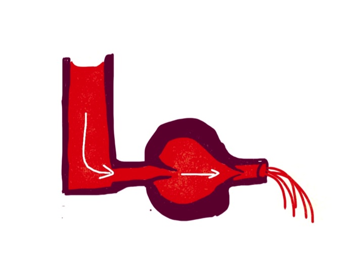
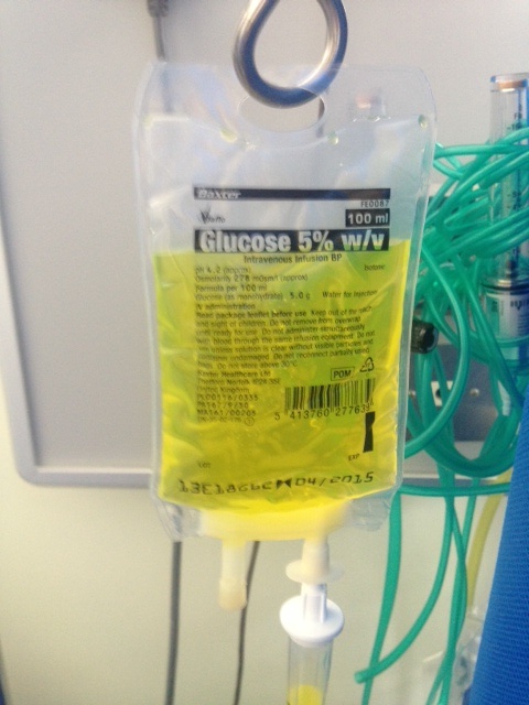
We took a blood sample and blood cultures, gave her intravenous morphine, paracetamol, gentamicin, co-amoxyclav and cyclizine (an antiemetic). She had pyelonephritis – a bacterial infection of her kidney.

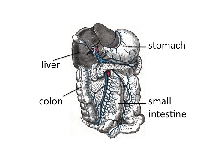
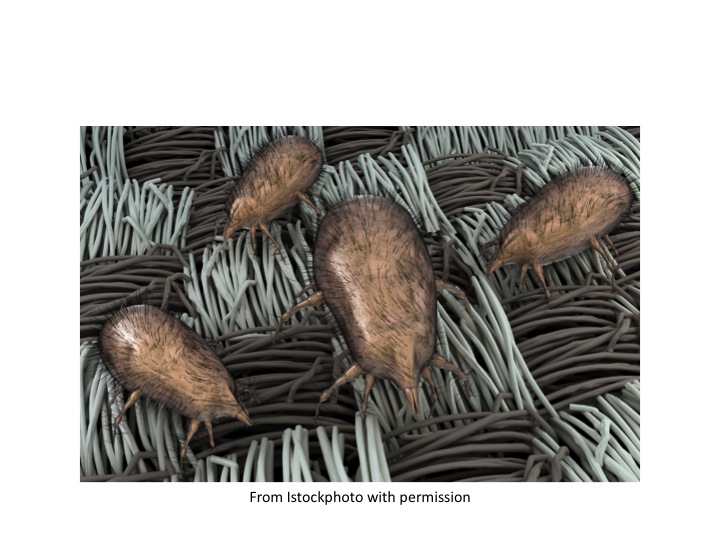
Young women get urinary tract infections much more commonly than young men. It’s to do with anatomy. The female urethra is very short, and germs can quite easily travel up to the bladder and then up the ureters to the kidney. Men get urine infections when they are older and have enlarged prostate glands.

Then they cannot empty their bladders completely and the stagnant urine is more likely to become infected. I was taught that urine is normally sterile in healthy people. It seems this is not the case. All of us have bacteria in our urine in small numbers – if you are interested read: http://www.ncbi.nlm.nih.gov/pmc/articles/PMC3744036/ 

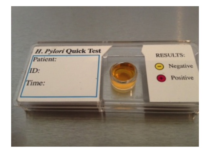
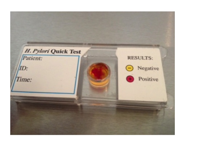
We use urine dipstick testing a lot on the acute medical unit. In theory it should be able to tell us which of our patients have a significant urinary tract infection. In practise it is not quite as useful as it should be. Kirsty’s urine tested positive for nitrites and leukocytes. In young women this is a good test, but we knew she had a urinary tract infection anyway. In elderly women, the tests are often positive even if a serious infection is not evident – perhaps because of the innocent commensal bacteria which are present.

Slide02

How do urine dipsticks work? The test for leukocytes is leukocyte esterase. In infected urine, the leukocytes are polymorphonuclear leukocytes, or neutrophils. I talked earlier (phlegm and horseradish) about how neutrophils, when they get excited by the presence of germs, make the enzyme myeloperoxidase which generates bleach. Esterase is another enzyme neutrophils make which breaks down peptide bonds, and specifically is useful in breaking down the peptidoglycan in bacterial cell walls. I guess its like getting stains out of clothes. Bleach works fine, but the proteolytic enzymes in washing powder can help too.

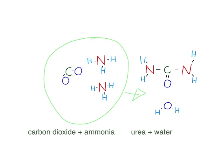
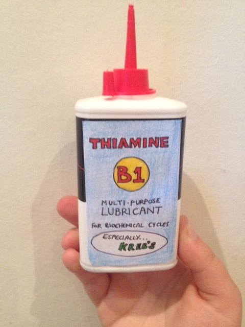
So what about nitrites? Bacteria like Eschericia coli, which are a common cause of urine infections are known as facultative anaerobes. This means that they can use oxygen to “burn” carbohydrates, protein and fats. But they can also use other “electron acceptors” to do this such as nitrate. Mammalian cells can only use oxygen. When I say mammalian cells, what I mean is mitochondria in mammalian cells – these small structures in our cells are responsible for all the energy generation from glucose, fats and protein. Think what happens when you eat a slice of toast. The amylase in our saliva starts to break down the starch in the toast to form glucose, a process which is finished by amylase in pancreatic secretions. This yields glucose, which is absorbed into the bloodstream. Glucose can be made into energy by the Kreb’s cycle, or citric acid cycle mainly happens in mitochondria. This is a complicated process, but essentially it means that glucose is turned into carbon dioxide and protons and electrons:

The mitochondria don’t get much energy from the Kreb’s cycle, but rely on the protons and electrons produced to make energy by combining them with oxygen. This happens in the electron transport chain:

Bacteria are more versatile than mammalian cells and can get energy out of these protons and electrons, even if oxygen is not available, by using nitrate instead of oxygen:

To do this they need to have the enzyme nitrate reductase. Human cells do not have nitrate reductase, so if nitrate is being turned into nitrite there must be bacteria present. So, if urine contains significant amounts of nitrite, the only way it can get there is if bacteria are using nitrate to “breathe” – a tell-tale sign that infection is present. The reason E.coli is called a facultative anaerobe is that it can survive by making energy if oxygen is present and also when it is not, by using molecules such as nitrate to “breathe”.

our mitochondria can only use oxygen to get energy from electrons and protons derived from glucose - some bacteria have nitrate reductase which can use nitrate as an electron acceptor - nitrite is a by-product- and appears in infected urine as an indicator of infection
our mitochondria can only use oxygen to get energy from electrons and protons derived from glucose – some bacteria have nitrate reductase which can use nitrate as an electron acceptor – nitrite is a by-product- and appears in infected urine as an indicator of infection

When I saw Kirsty later in the afternoon she was much better. The pain had not gone, but was eased by the morphine. Her temperature had come down, and she had stopped vomiting. Her infection was coming under control.

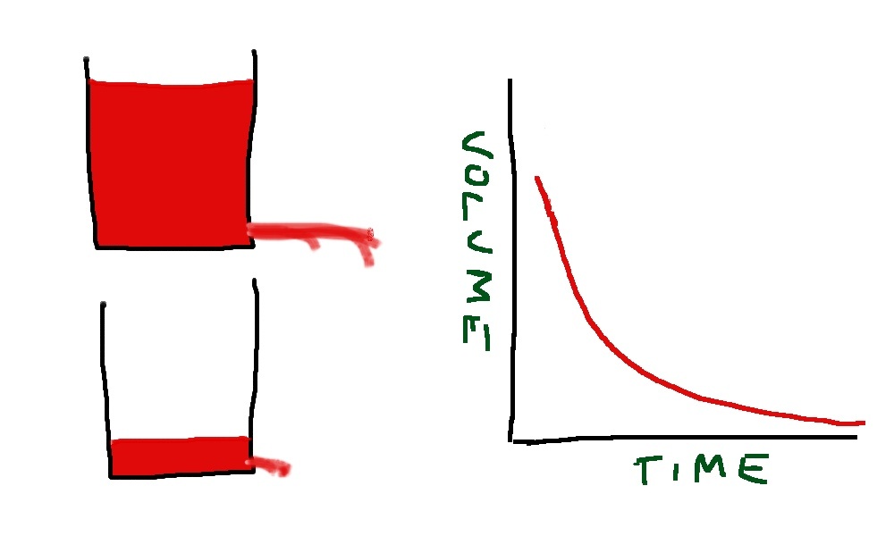
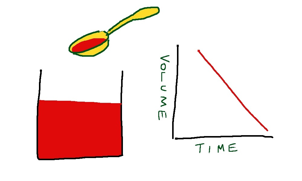
Both gentamicin and co-amoxyclav are very effective in treating urinary tract infections. They are both rapidly excreted by the kidneys and achieve much higher concentrations in the urine than in the bloodstream. Gentamicin has a half-life of about 2 hours in patients with normal renal function. So, lets say we give a person 250mg of gentamicin intravenously. The blood volume is about 5 litres, so the immediate concentration will be 50mg/litre of gentamicin. In two hours about 100ml of urine will be made, and half of the gentamicin previously given intravenously will be in that urine. That is 125mg in 100mls or 1250mg/litre – more than twenty times the concentration in blood. Amoxycillin has a half-life of more like one hour, so achieves even higher urine concentration in comparison to blood.

What do gentamicin and co-amoxyclav do? They are antibiotics that work in quite different ways.

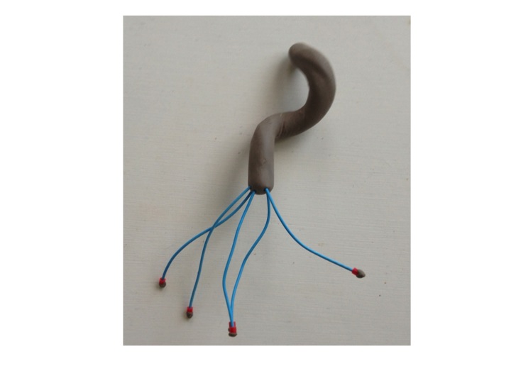
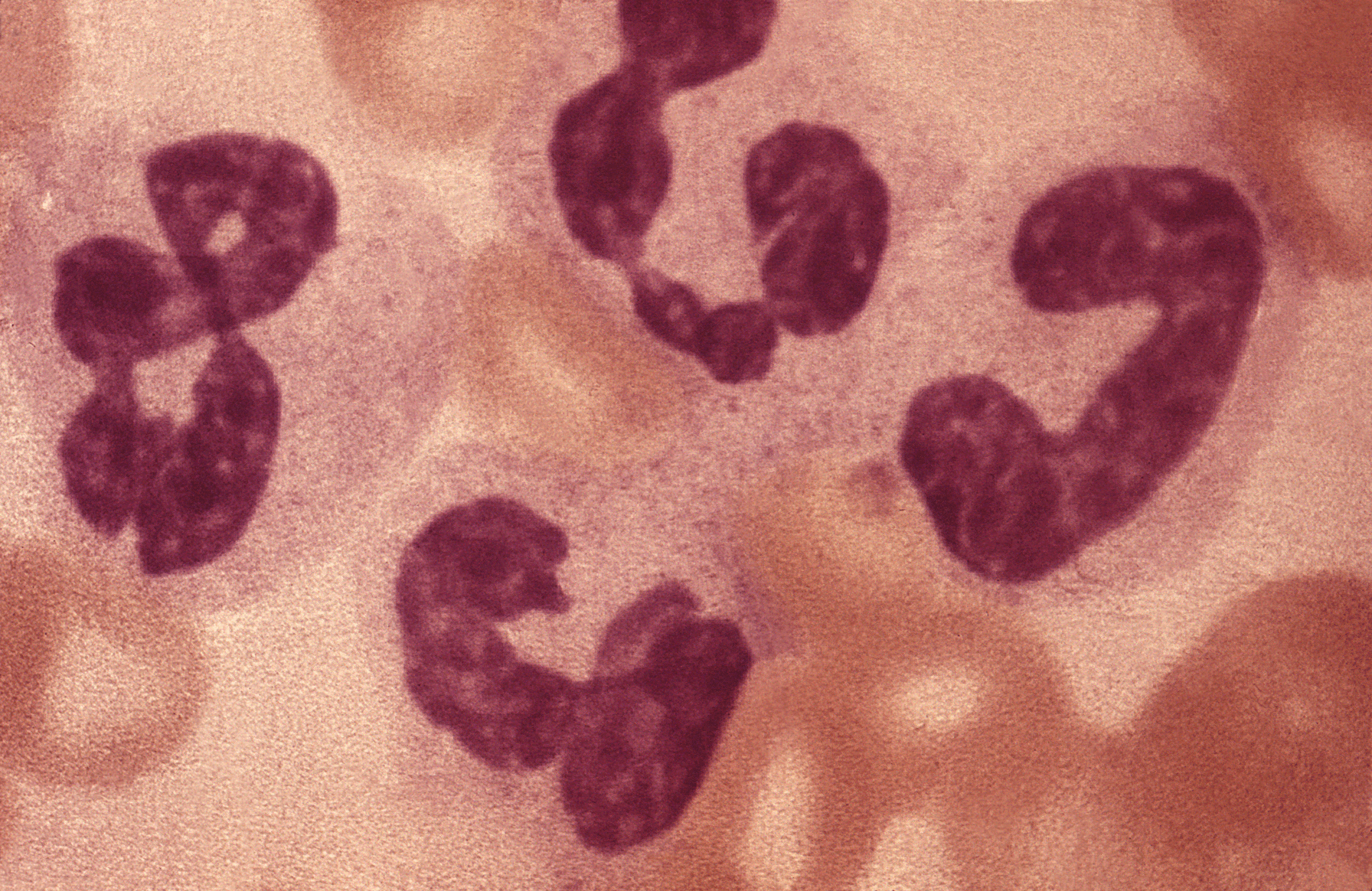
Gentamicin is an aminoglycoside. That means it is a sugar with amine groups. Here is the structure – just three sugars with lots of NH2 groups:

gentamicin is a relatively small molecule with three sugar groups and lots of amine groups (in red) - an aminoglycoside
gentamicin is a relatively small molecule with three sugar groups and lots of amine groups (in red) – an aminoglycoside

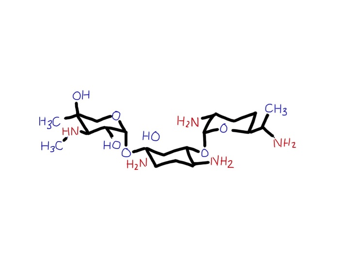
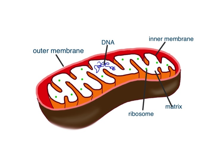
It gets into the bacteria and binds strongly to its ribosomes. These are the really important and clever machines in bacteria which make proteins from DNA. Mammalian cells also have ribosomes to make our proteins – but they are not at all the same. They work in the same way but over the past 2 billion years have changed with evolution so they are a different shape and are larger than bacterial ribosomes. Gentamicin does not interfere with mammalian ribosomes. For an antibiotic to be useful it has to damage bacteria but not human cells. Luckily, ribosomes are so different between bacteria and mammalian cells that some chemicals such as gentamicin will selectively bind only to bacterial ribosomes.

mitochondrion - it has all the stuff inside that a bacterium has, but without a tough cell wall
mitochondrion – it has all the stuff inside that a bacterium has, but without a tough cell wall – redrawn from wikipedia – author kevinsong

Other antibiotics such as tetracyclines, macrolides (erythromycin and clarithromycin), chloramphenicol and clindamycin also work by interfering with bacterial ribosomal function. A bacterium with damaged ribosomes has major problems – it cannot make new proteins. That means it cannot divide and make new bacteria. It will be immobilised and suffer a slow and painful death. (Not really, I don’t think bacteria don’t feel pain – but then I don’t have evidence for that). If it is a bacterium which makes a protein toxin, such as staphylococcus causing toxic shock syndrome, turning off protein production with a ribosomal poison such as clindamycin is a good idea – rather than causing bacterial cell wall damage and leakage of more toxin with penicillin therapy.

Gentamicin can cause problems if it is given over prolonged periods, because it can accumulate and cause damage to ears and kidneys. The damage to hearing is probably due to damage to mitochondria. More specifically damage to mitochondrial ribosomes. We only gave Kirsty one dose of gentamicin – problems with this drug usually happen when patients with impaired renal function are given aminoglycosides for several days, or when aminoglycosides are given with other drugs such as vancomycin which can impair renal function.

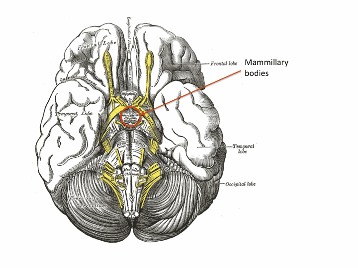
Mitochondria are thought to derive originally from bacteria. Once upon a time, a long time ago there was a cell that survived well enough by getting energy from glycolysis – turning glucose into pyruvate. This cell did not need any oxygen. It scraped a living producing at most 2 ATP molecules per glucose molecule. Then it had a conversation with a bacterium which said “Hi doll, I could take that pyruvate you make and turn it into another 28 ATP molecules by combining it with oxygen – how about it?” Maybe this conversation happened at the time oxygen had begun to appear in the atmosphere (see great oxygenation event in last week’s blog). “With your looks and my talent we could do Broadway together”. This is technically known as endosymbiosis, where one type of cell engulfs another to work together to their mutual benefit. The result is eukaryotic cells – the cells we are made of. Our cells contain mitochondria that derive from bacteria. They can make lots of energy from glucose and oxygen. The bacteria are looked after and nurtured inside the cells which engulfed them. Like bacteria, these mitochondria have their own ribosomes, that, not surprisingly, are similar to the ribosomes of bacteria that are causing Kirsty’s pyelonephritis. Too much gentamicin can damage mitochondrial ribosomes and cause hearing loss – see:

Click to access 9443888.pdf

We also gave Kirsty co-amoxyclav.

the basic penicillin molecule - the central beta lactam is in the pink circle
the basic penicillin molecule – the central beta lactam is in the pink circle

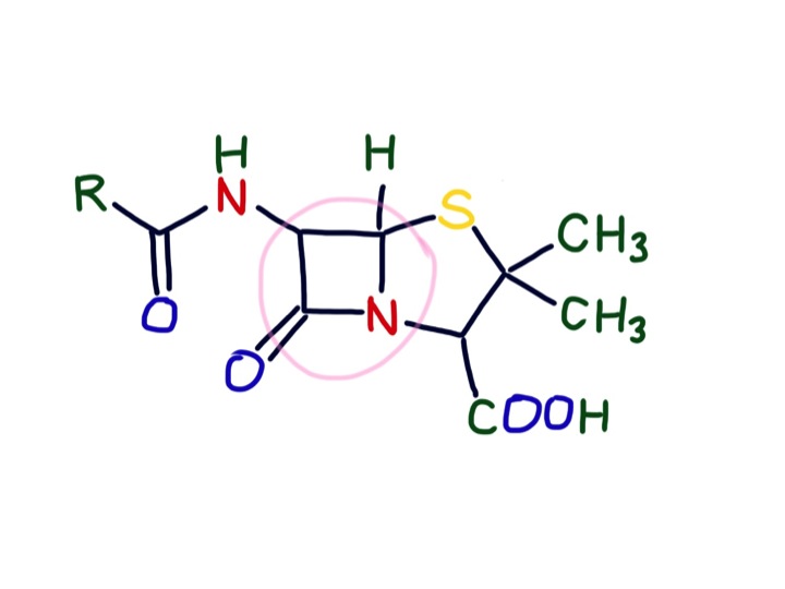
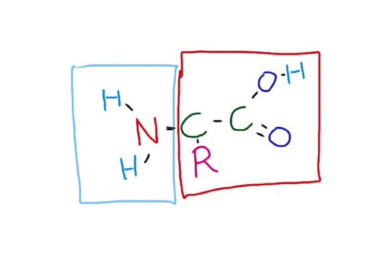
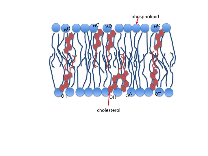
This is a combination of amoxicillin and clavulanic acid. Amoxycillin is a penicillin. Originally discovered by Alexander Fleming, the original penicillin, benzylpenicillin, has been modified by pharmaceutical companies to be more effective. Unlike benzylpenicillin, amoxycillin is rapidly absorbed by the stomach. It is also effective against gram negative organisms such as E.coli. Penicillins have a beta lactam group. This structure makes it difficult for bacteria to make a vital component of their cell wall – peptidoglycan.  This is a tough polymer made of special sugars and short peptide chains. The beta-lactam group in penicillins is the right shape to get stuck in the cell wall building enzymes and prevent cell walls being made. Our cells do not have cell walls – they just have thin, delicate plasma membranes made of phospholipid and cholesterol. Similarly, although mitochondria are similar to bacteria, they also do not have cell walls. Mammalian cells and their mitochondria are very cosseted and protected in a 5-star luxury apartment with all mod-cons.  They are looked after in a temperature-controlled environment. Oxygen is supplied free and waste carbon dioxide and other unwanted substances taken away continually. Acidity is tightly controlled – pH between 7.35 and 7.45, osmolarity not too high or low. The poor bacterium, in contrast, has to tolerate acid, alkali, high and low osmolarity and a whole host of chemical insults as well as having to find its own food. And then there is the danger of being chased by an angry green neutrophil. No wonder it feels happier with a thick, tough cell wall to protect it. The clavulanic acid is to inactivate beta lactamase – an enzyme some wily bacteria have started making to destroy beta lactam antibiotics. No doubt the bacteria will soon be making betalactamaseinhibitorase enzymes.

There are other ways to selectively attack bacteria without harming human cells. All cells need folate to manufacture nucleic acids. We get our folate from diet – particularly green, leafy vegetables (folate is related to the word foliage). Bacteria do not, in general, have a healthy diet. They instead make the large folate molecule themselves from much smaller molecules. Trimethoprim and sulphonamides prevent bacteria from making folate causing them to suffer and die a “thymineless death” (look it up in Wiki).

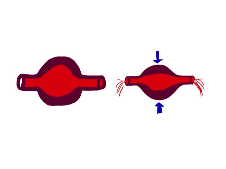
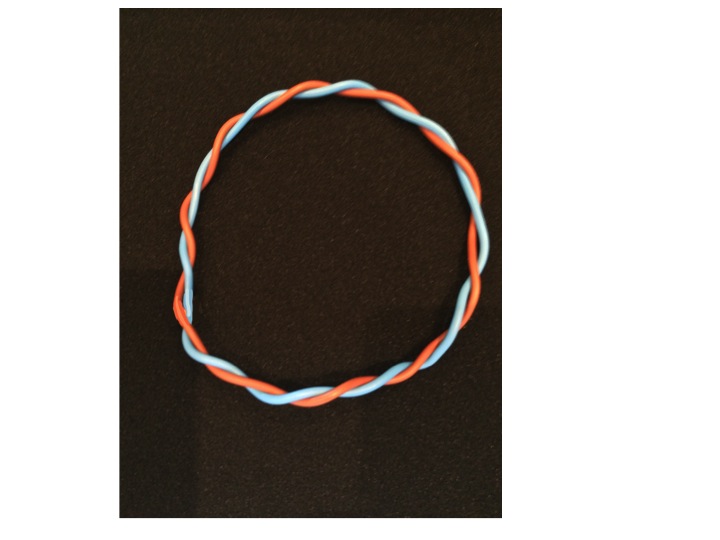
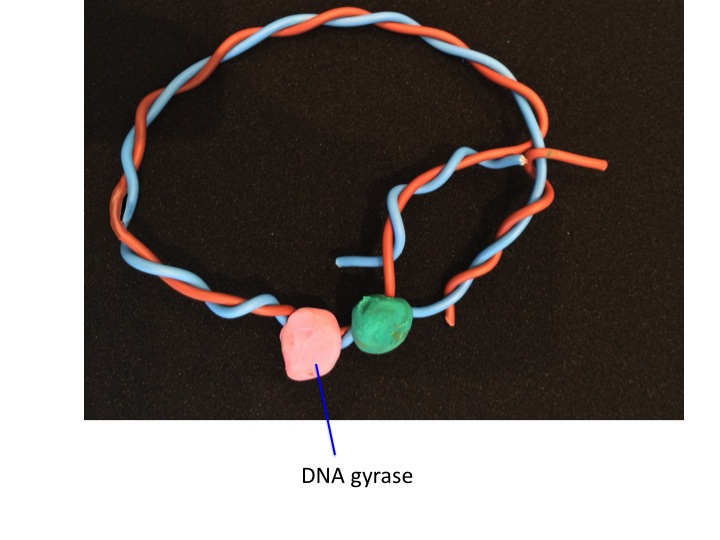
Fluoroquinolones such as ciprofloxacin are some of the newer antibiotics which have come into clinical use since I qualified. They were hailed as the new wonder drug, but we now use them relatively rarely because they particularly seem to promote C.difficile infections in frail, elderly people. They work by inhibiting DNA gyrase and Topoisomerase IV. I hope the illustrations will explain how they work:

bacterial DNA is circular
DNA helicase pulls the DNA strands apart so that they can be replicated to make more DNA  when the bacterium divides - but it causes a problem, the DNA becomes supercoiled
DNA helicase pulls the DNA strands apart so that they can be replicated to make more DNA when the bacterium divides – but it causes a problem, the DNA becomes supercoiled
DNA gyrase sorts out this problem - cutting the DNA strand and rejoining it having removed the twist
DNA gyrase sorts out this problem – cutting the DNA strand and rejoining it having removed the twist – fluoroquinolones such as ciprofloxacin stop this enzyme working
having replicated the DNA - the two circular strands are interlinked!
having replicated the DNA – the two circular strands are interlinked!
but topoisomerase IV comes to the rescue and chops the chain and rejoins it to separate the circular strands
but topoisomerase IV comes to the rescue and chops the chain and rejoins it to separate the circular strands- fluoroquinolones also inhibit this enzyme
all sorted
all sorted

The food link this week is blue cheese.

blue stilton
blue stilton – the blue bits are penicillum mould

The reason it is blue is because of the growth of the mould penicillium, which is a grey/greenish blue colour.  The spores are blue, not the fungus itself.

volkornbrot past its sell by date - the blue mould is penicillium - not sure what the yellow stuff is - any ideas anyone?
volkornbrot past its sell by date – the blue mould is penicillium – not sure what the yellow stuff is – any ideas anyone?

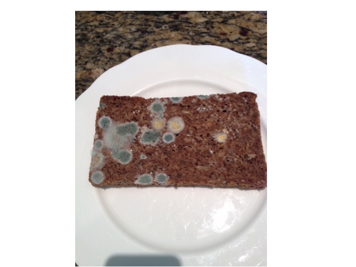
The mould in Roquefort and Stilton is P. roquefortii, a close relative of P. notatum (now known as P. chrysogenum), the penicillium mould that Alexander Fleming found inhibiting the growth of staphylococci.

I’ll be in France next week so the next post will be in 2 weeks’ time

vomiting blood and yoghurt

Slide04 More alcohol problems this week. Peter came in vomiting blood yesterday afternoon. Vomiting blood is always a bad thing, but this time it was particularly bad. His long-suffering partner Rita came in with him to tell us what had happened. Peter, aged 69 was too drowsy and confused to tell the story himself. He was a retired barman and had always drunk too much alcohol. He recently went to see the liver doctors because his abdomen had filled up with fluid. They told him that he would die soon if he did not give up drinking. Rita said that he had cut down but was still drinking about 3 pints of strong cider every day. Yesterday lunchtime he was about to sit down to eat when he said he felt very sick. He staggered to the bathroom and promptly vomited what Rita estimated to be a pint of bright-red blood down the toilet pan. Rita called the ambulance. He vomited more blood on the way and by the time they arrived he was pale, sweaty and quite drowsy. She was really worried – she thought about what the liver doctor had said about Peter dying soon if he did not give up drinking – he had not given up.Slide05

When patients with liver disease vomit blood it always makes us worry about bleeding varices. Varices are large, distended veins which appear at the junction between the oesophagus (foodpipe) and stomach in people with liver cirrhosis. Cirrhosis often results from liver damage due to alcohol. Ethanol is metabolised to ethanal (acetaldehyde) and causes damage to liver cells as well as the pancreas (see vodka and sweetbreads below). The liver does have a remarkable capacity to regenerate.

Slide06

I’m not sure if the ancient Greeks knew about liver regeneration when they devised the myth about Prometheus. He made the mistake of giving fire to men, and as a punishment was chained to a rock for eternity. Every morning an eagle would fly down and peck out his liver. During the following day his liver would grow back again, to be pecked out again the following morning. He is still there.

ancient Greek vase showing Prometheus having his liver pecked out by eagle wikimedia common user Bibi Saint-pol
ancient Greek vase showing Prometheus having his liver pecked out by eagle –  wikimedia common user Bibi Saint-pol

Although liver does regenerate when damaged by alcohol, it does so to form nodules of liver tissue with bands of fibrosis in between the nodules. This disturbance of normal architecture impairs blood flow through the liver. As a result the pressure in the portal veins carrying blood from the stomach and intestines to the liver increases. Increase in portal venous pressure results in oesophageal varices. When they burst, rapid death from blood loss is common. Another result of increase in portal pressure is ascites – fluid accumulation in the abdominal cavity – the cause of Peter’s abdominal swelling.

all the blood coming from the stomach and small and large intestine goes into the portal venous system and through the liver to be processed - including removal of ammonia and small amines
all the blood coming from the stomach and small and large intestine goes into the portal venous system and through the liver to be processed – including removal of ammonia and small amines – from Grays anatomy 1918

So he was filled up with a blood transfusion, vitamins (see vodka blog below) and given terlipressin – a drug which constricts oesophageal varices and helps to stop bleeding. He was sent as an emergency to have an upper gastrointestinal endoscopy. In fact he did not have significant varices. He had a bleeding duodenal ulcer. The ulcer was cauterised and injected with adrenaline, a biopsy was taken from his duodenum, and he was sent to the admissions unit.  Part of the duodenal biopsy was put into a CLO test kit.

I talked previously about adrenaline causing muscle tremor and relaxation of bronchial smooth muscle by activating adrenergic beta receptors. It is released by the middle (medulla) of adrenal glands in response to severe stress. The reason Peter was so pale was probably more to do with release of adrenaline than blood loss. This hormone has many other actions to help us survive life-threatening situations. It will also act on beta receptors in muscle blood vessels to increase muscle blood flow – good to get away more quickly from the nasty tiger with dripping fangs that likes to eat humans.

Skin blood vessels have few beta receptors – here adrenaline acts on alpha receptors to cause reduction in blood flow. Similarly, in the lining of the duodenum, adrenaline, when injected by the endoscopist, causes blood vessels to constrict and help stop bleeding by acting on their alpha receptors.

When I ask students why adrenaline reduces skin blood flow they usually say it is to redirect blood to the central circulation where it is more needed. The skin only has about 1% of circulating blood. It is more likely that skin blood flow is reduced to limit blood loss when the tiger’s teeth finally sink into that tasty human flesh.

All this preamble is a good excuse to talk about urea. In our hospital normal blood urea levels are between 3.5 and 6 mmol/l. In the US doctors talk about blood urea nitrogen- the same stuff- normal levels are 20-30mg/l. When Peter visited the liver doctor last month his blood results showed that the concentration of urea in his blood was low – only 1.8 mmol/l (5mg/dl BUN). When he arrived in the ED it was elevated at 14 mmol/l (40mg/dl BUN). His haemoglobin was low at 90g/dl and clotting was deranged with an INR of 2.9. Why was his urea low before and now high? To understand this I need to talk about protein metabolism.

Most of us in the West eat lots of protein. More than we need. In the US and UK adults eat about 100grammes of protein/day, although we only need about 50. If we eat 100grammes of protein a day, we need to get rid of the same amount, unless we are growing, body-building or pregnant. Patients who are ill typically break down more protein than they take in – negative nitrogen balance.

I’ve used arguments about in/out balance for fluid in my previous post and will use it for energy in future posts. Not everything in humans can be understood in terms of in/out balance. For instance, with my 11 year-old son we input high grade educational material and all that comes out is poo and fart jokes.

So what happens to this 100grammes of protein? Protein is a polymer of amino acids. You can make useful plastic out of milk protein – see this youtube video:

http://www.youtube.com/watch?v=pIvAl4lu1uA

Casein also used to be used to make plastic items such as buttons. We now have cheaper and better plastics.

Protein that goes into our mouth is mashed up by our teeth and swallowed. The stomach has the first go at breaking up the protein polymer with the enzyme pepsin, secreted by chief cells in the glands of the stomach. It’s a bit tricky making an enzyme that breaks down protein, because all our cells are made of lots of proteins.  There is the obvious danger that the enzyme will destroy the cell that made it. So pepsin is made in an inactive form – pepsinogen that is only activated when it comes into contact with stomach acid. Then the pancreas has a go. It makes trypsin, carboxypeptidase and chymotrypsin which finish the job, to end up with amino acids. The pancreas has to be pretty careful too, making inactive enzymes which become active one secreted – see:

http://www.physiologymodels.info/digestion/proteins.htm

Amino acids are all of the general formula:

general formula for amino acid - the red box stuff can usually be turned into carbon dioxide and water - the blue box stuff is harder to get rid of
general formula for amino acid – the red box stuff can usually be turned into carbon dioxide and water – the blue box stuff is harder to get rid of

The R is mainly made of carbon, hydrogen and oxygen. We have to dispose of 100g daily. The part in the red box is burned up in mitochondria, and like glucose is turned into carbon dioxide, water and energy – 4Kcal/gramme – 400 Kcalories per 100g. The problem is the bit in the blue box. The NH2 group looks like ammonia, and ammonia is toxic. But don’t worry, we have a way of dealing with this – it’s called the urea cycle. This happens mainly in the liver. I could draw a diagram of the urea cycle, but instead will direct you to a 1 minute youtube video showing how it works:

http://www.youtube.com/watch?v=AoBbVu5rnMs

So ammonia is combined with bicarbonate (or carbon dioxide) and via the urea cycle is made into urea, a very non-toxic substance which is excreted in urine.

two molecules of ammonia combine with one of carbon dioxide to make urea and water - its not as simple as that - but that is what the urea cycle does
two molecules of ammonia combine with one of carbon dioxide to make urea and water – its not as simple as that – but that is what the urea cycle does

The reason Peter’s urea was so low when he saw the liver doctor was because he was drinking too much alcohol and not eating enough protein. Now he has had a sudden high protein meal – blood. Blood contains about 70g/l of albumin and globulins in plasma, and about 130g/l of haemoglobin (mostly protein) in red cells – total 200g/l.  If he has bled a litre of blood from his duodenal ulcer he will suddenly have twice the average daily protein intake in a short time – no wonder his urea level has risen.

But Peter’s liver is not working as well as it should, because he has been drinking too much alcohol for a long time and has cirrhosis. A lot of the ammonia from protein metabolism is not being instantly turned into urea, and instead gets into the systemic circulation. When the brain is exposed to ammonia, it does not function too well. It is not only ammonia, but other short-chain amines which the liver has failed to deal with. Bacteria in the colon chop up amino acids and proteins into all sorts of amine-containing molecules which the liver will normally cope with. Peter’s damaged liver is not able to cope with these substances and they get into his circulation and into his brain. Nobody knows how brains work, but we do know that certain chemicals are important in passing messages from one brain cell to another – neurotransmitters. Many neurotransmitters are small amine-containing molecules, such as catecholamines, serotonin (5-hydroxytryptamine), glutamine and dopamine. It is not surprising that when flooded with a smorgisbord of small amines and ammonia the brain does not work too well. Rita told us that Peter was sleeping all day and awake all night recently – a characteristic feature of hepatic encephalopathy. When we examined him when he came back from endoscopy he had a liver flap, or asterixis. That means when he held out his hands they would independently twitch, with a downward flapping movement- another feature of hepatic encephalopathy which was also making him drowsy. There are lots of examples of liver flap on youtube such as:

http://www.youtube.com/watch?v=Rbv-zaVszlk

The reason his INR was raised was because his liver was not producing enough clotting factors. We gave him a concentrate of clotting factors (octiplex) to correct this, and help stop the bleeding.

It took a couple of hours before his CLO test result was available. A small piece of tissue was removed from his duodenum next to the ulcer. The CLO test refers to Campylobacter Like Organism. Helicobacter pylori is one of these which is a major cause of duodenal ulcers. Helicobacter pylori is an amazing germ. It is a bacterium which can survive in a very hostile environment – the human stomach and duodenum. It is the only germ that can normally survive here. The acidity is fierce, often the pH is down to 1 – really strong hydrochloric acid. There is also a high concentration of nitric oxide and proteolytic enzymes. It doesn’t mind.  Helicobacter refers to the shape – a helix or corkscrew.

model of helicobacter showing corkscrew shape and long flagellae

I talked before about how bacteria find it difficult to swim in mucus. The stomach lining is covered in mucus, but helicobacter’s corkscrew shape helps it swim in this thick and gloopy layer. Staying in the mucus layer helps protect it from the acid and enzymes. Another protection comes from being able to convert urea into alkaline ammonia – keeping the acidity at bay in its immediate environment. The CLO test simply tests the small bit of duodenum for its ability to convert urea into ammonia – it can only do this if has helicobacter organisms in it. Human cells cannot do this. So, in the test plate, there is an indicator, such as phenolphthalein or phenol red which changes colour with alkali.

this CLO test kit contains phenol red - it is yellow when acid and red when alkaline
this CLO test kit contains phenol red – it is yellow when acid and red when alkaline

The helicobacter turns urea into ammonia, making the environment more alkaline which will change the colour of the indicator. Phenol red changes from yellow to red with alkali.

when a piece of duodenum is put into the CLO test vial it will turn red if there are helicobacter pylori organisms - I just used a bit of soap for this photo as I did not have any infected duodenum to hand
when a piece of duodenum is put into the CLO test vial it will turn red if there are helicobacter pylori organisms – I just used a bit of soap for this photo as I did not have any infected duodenum to hand

We gave Peter intravenous omeprazole, a proton pump inhibitor. This stops the parietal cells of the stomach making acid and helps heal the ulcer. We also gave him lactulose. This is a sugar which helps with hepatic encephalopathy. Lactulose is very similar to lactose – the sugar in milk. Lactose is a disaccharide made of the simple sugars glucose and galactose. Lactulose is made of fructose and galactose. I talked earlier (chest pain) about how most of the world’s population have problems drinking milk when they are adult because they have lost the enzyme which breaks down the bond between the two sugars in lactose. No-one has the enzyme to break down lactulose into its two simple sugars. But bacteria do. In the large intestine, lactulose that we have been unable to break down and absorb in the small intestine is turned into lactic acid, which is also then turned into lots of gas, such as methane and hydrogen. Some patients don’t like lactulose because it gives them stomach cramps and makes them fart a lot. But in patients like Peter it is good because the lactic acid reacts with the alkaline ammonia and small amines to inactivate them and reduce their amount in the circulation affecting his brain, making him confused and drowsy. He went home less confused and more or less OK. I hope he stops drinking.

This is where the yoghurt comes in. The food link at last. In yoghurt the milk sugar lactose is broken down by lactobacilli to form lactic acid. This gives it a nice tangy taste that my daughter likes so much she wears the following tee-shirt.Slide08

heart failure and haggis

Heart failure is a very common reason to be admitted to our hospital. John, age 73, had a very typical story. He was well until about 3 years ago when out of the blue he had a major heart attack. Life has never been quite the same since. He has to take lots of tablets – aspirin, statins, beta blockers, ACE inhibitors and several fish oil capsules.

His wife worries about John a lot. She no longer makes him steak and kidney puddings and has stopped him smoking cigars when he goes out with his friends on a Saturday night. He has bought a dog and now takes much more exercise and drinks a lot less beer. But life wasn’t too bad until he noticed he was getting more short of breath when he took the dog for a walk a few weeks ago. Then he noticed ankles were swelling – his socks would leave deep rings around his ankles which disappeared in the morning. For the past week he found he could not lie flat without feeling short of breath.

Then the night he was brought into hospital he woke up gasping for breath. He was pale and sweaty and his wife got really worried and called the ambulance. By the time the paramedics got there he was still quite breathless and his ECG was abnormal, so they brought him in to hospital. He was given some nitroglycerin spray under his tongue- this helped a lot. In the emergency department he was given a dose of intravenous furosemide that made him “pee for Britain”.

We examined him on the admissions unit and found he still had some swelling in both his ankles which left dents when squeezed – bilateral pitting oedema- what was once known as cardiac dropsy. His neck veins were fuller than normal and he still had crackles at the base of his lungs when we listened. He had pulmonary oedema due to heart failure.

I really don’t like the term heart failure. You will know that I’m not one for using long Latin words but something like “impaired cardiac function” might be a less alarming label.

So why did John’s ankles swell up and why did he become breathless? When I ask students and junior doctors they usually say something about back pressure and right heart failure. Maybe it’s just in the UK, but I suspect there is a lot of confusion and misunderstanding about the physiology of heart failure. Pulmonary oedema is about back-pressure, but raised central venous pressure and peripheral oedema is not. So what causes ankle swelling in heart failure? What follows is my understanding of what is a complicated problem. I’ll start slowly and it may take a while to get to the answer. This is the most difficult thing I’ve tried to explain so far.

First I want you to imagine a simple pump. The pump is a hollow rubbery container which works because it is made of muscle which contracts rhythmically. At each contraction the volume in the chamber is much less than when it is relaxed. For a normal left ventricle about 60% of the blood is squeezed out each time it contracts (ejection fraction).

the ventricles are made of muscle which squeezes fluid - without valves it would not pump in one direction
the ventricles are made of muscle which squeezes fluid – without valves the heart would not pump in one direction

To make the blood go only in one direction, it is important to have valves at the inlet and outlet of the pump. There are three things helping to fill the pump when the muscle is relaxed. There needs to be a filling pressure, represented in the diagram below by the height of fluid in the left-hand reservoir. In the left side of the heart this is pulmonary venous pressure, in the right it is central venous pressure, most easily measured by looking at the height of blood in the neck veins. But lets first of all think about a simpler system with only one pump and one filling reservoir. Many years ago Frank and Starling discovered that if the filling pressure of a heart chamber affected how well it pumped blood out. The higher the filling pressure the stronger the contraction, making the pump work harder.

the higher the column of blood in the reservoir, the harder the heart pumps - it now has valves so the blood can only go in one direction
the higher the column of blood in the reservoir, the harder the heart pumps – it now has valves so the blood can only go in one direction

A lot of hollow things in our body work in a similar way. The uterus starts to contract when it gets stretched enough by the increasing size of the unborn baby. Our intestines work much harder if they are blocked, causing the wall to be stretched – this results in colic. The same is true for the gallbladder and ureters- both painfully contract when a stone causes a blockage and the muscular wall is stretched.

The second factor helping our heart chambers to fill up is suction. A turkey baster bulb will suck up fluid because the rubbery stuff it is made of wants to spring back to its normal shape. The same is true for the ventricles of the heart – they actively suck fluid in while relaxing during diastole. I am told that if you put an isolated animal heart into a bucket of oxygenated physiological saline solution it will move around like a squid – sucking liquid in and squirting it out to propel it along in the fluid. It could only do that if the ventricle actively sucks in fluid.

The third important factor in ventricular filling is the contraction of the atria. These give a bit of extra stretch to the ventricles by pumping in some more blood  to help encourage them to pump it out a bit harder.

The heart pumps blood around a circuit – the circulation. It first goes into the aorta, but the vessels get progressively smaller in diameter, ending up with capillaries which have an internal diameter of less than that of a red blood cell – about 7 microns.

capillaries have just one layer of cells and are just big enough to allow a red blood cell through

Pushing blood through the large vessels is very easy, but much more difficult through the smaller vessels. There is a strange relationship between the amount of resistance to flow and the internal diameter of a pipe which was discovered by Pouseuille- resistance is inversely proportional to the fourth power of the internal diameter. In practice this means that most of the resistance to flow is in small vessels with a hole down the middle measuring  less than one tenth of a millimetre – these are also known as resistance vessels. Because of Pouseuille’s law, resistance vessels can constrict or relax a very small amount to cause a large change in flow to any particular organ – depending on its requirements for oxygen.

Back to the circuit. The arteries are thick-walled, with a relatively small hole down the middle. Veins are thin walled and can accommodate more volume. So most of the blood in our circulation is in the veins. Blood is pumped into the large arteries, is squeezed through the resistance vessels, and then ends up in the veins. You will not be surprised to know that the amount of pressure in the large arteries depends both on how hard the pump is pumping – cardiac output, – and how much resistance to flow there is in the small arteries – peripheral vascular resistance. Pressure = Flow x Resistance.

One thing our bodies really care about is the pressure in our large arteries – commonly known as blood pressure. It is continually monitored by pressure-sensing devices in the aorta and carotid arteries. These baroreceptors send messages to the brain stem (medulla). If the pressure is low, the medulla sends messages via the sympathetic nervous system to the heart to make it pump harder, and to the resistance vessels to make them constrict a bit to get the pressure back to normal. The medulla also sends messages to the veins to make them constrict – we will see why in a moment.

Looking at our simple circuit, blood pressure is determined by how hard the heart is pumping, and how much resistance to flow there is in small vessels. What determines the pressure on the venous side – the filling pressure of the reservoir (on the left side in the diagram)?

blood moves around in a circuit - the pressure (height of column of blood) in the reservoir will not change if the pump is pumping fast or slowly - but the pressure on the arterial side will very much depend on how much the pump is pumping
blood moves around in a circuit – the pressure (height of column of blood, or central venous pressue) in the reservoir will not change if the pump is pumping fast or slowly – but the pressure on the arterial side will very much depend on how much the pump is pumping

If the heart pumps less hard, or the resistance changes – I hope you can see that neither of these things will make a difference to filling pressure in the reservoir in this simple system. When students talk about “back pressure” they seem to forget that the blood goes round in a circuit. Failure of any part of the heart will not, on its own, affect filling pressure of the right side of the heart. Central venous pressure is determined by only two things: the amount of fluid in the system and how much veins are constricted. The amount of fluid in the circulation is affected by (a) how much fluid we drink every day and (b) how much we lose. Most of the fluid loss is in the form of urine (see previous post). It is the job of our kidneys to regulate the volume of blood in our circulation. Normally they do a fabulous job. If I drink a pint of beer, half an hour later I will be needing to have a pee – lots of dilute urine has been produced to get rid of all the extra water. If I eat two packets of crisps with the beer, over the next few hours my kidneys will get rid of the extra salt with no problem. Kidneys work in a timescale of hours.

central venous pressure is very much affected by the volume of blood in the circulation, which in turn depends on the balance between fluid input and output - cups of tea and daily urine volume
central venous pressure is very much affected by the volume of blood in the circulation, which in turn depends on the balance between fluid input and output – cups of tea and daily urine volume

Venous constriction works in a timescale of seconds. If I am in a chair and about to get up and walk around, my brain stem will send messages via the sympathetic nervous system to my veins to tell them to constrict. Squeezing this complex network of floppy tubes in my legs, belly and chest will increase cardiac filling pressure and, as Frank and Starling discovered, increase the force of contraction of my heart to supply my muscles with more oxygen. Understanding of how heart failure causes problems needs an understanding of how kidneys and veins work, not just the heart.

Now I am going to make the circulation model a bit more complicated. We have two pumps, not one. They are joined in series. The right heart pumps blood into the lungs. It returns to the left heart which pumps it round the rest of the body. The diagram shows the two pumps separated, but of course in real life they are part of the same organ.

this diagram shows the two sides of the heart separated - the pressure in the pulmonary vein is higher than the central venous pressure on the right side
this diagram shows the two sides of the heart separated – the pressure in the pulmonary vein is higher than the central venous pressure on the right side

The fact that they are arranged in series clearly could lead to problems. What if the right heart tries to pump more than the left? Why is this not normally a problem? The way the system is set up is that the right heart pumps blood through the lungs into the pulmonary veins. Here the pressure is higher than the normal 7cm water central venous pressure – in health about 16cm water. This higher pressure in the pulmonary vein is useful priming device so the left ventricle, which is bigger and stronger, can be rapidly filled and has plenty of pressure to stretch the rubbery myocardium to enable it to contract hard and generate adequate systemic arterial blood pressure. When right heart output increases, the pulmonary venous pressure will temporarily increase, and left ventricular output will in turn increase because of the Frank-Starling law, and subsequent reduction of pulmonary venous pressure to normal. The end result is that the output of the left side of the heart matches that of the right, and pulmonary venous pressure has not changed much – all sorted. This does mean, if you think about it, that the cardiac output is being controlled by the right heart – it pumps what it wants and the left heart has to follow suit. It is not always the case that the bigger, stronger partner is in control.  John has found that out recently.

What controls right heart output? Well the brain stem has a big say, by constricting veins and increasing filling pressure and by sending messages to the heart via sympathetic nerves. But the kidneys are also really important – by altering fluid balance they can control filling pressure. Kidneys are involved in turning the dials and can really mess things up when they get it wrong, as we shall see.

Lets get back to John. His left ventricle was damaged by a sizeable myocardial infarction a few years ago (see chest pain and horsemeat lasagne below). Soon after had an echocardiogram which showed that the part of his left ventricle was not contracting as well as it should – the overall ventricular ejection fraction was found to be 44%.  It was probably about 60% before. The rest of his left ventricle which was not damaged has to work harder to maintain his cardiac output and blood pressure. Heart muscle is different from ordinary skeletal muscle – the sort which makes our arms and legs work. Skeletal muscle responds very well to being worked hard. It grows bigger and stronger to more exercise we take. The heart muscle does this to an extent, but eventually it seems to give up and stop working so well. I don’t think anyone quite understands why this is. If experimental animals are infused with isoprenaline, a drug which makes the heart beat strongly by stimulating heart muscle cells in a similar way to noradrenaline released by sympathetic nerves, their hearts will fail in about 2 weeks. Beta blockers stop this sympathetic stimulation, and patients with heart failure live longer if they take them regularly.

Poor cardiac output stimulates the kidneys to produce renin. This is an enzyme which generates angiotensin I. Conversion of angiotensin I to angiotensin II on the surface of blood vessels helps restore blood pressure but also has a bad effect on heart muscle. It seems to make the muscle cells produce damaging free-radicals and oxidising agents which cause cardiac muscle death. ACE inhibitors prevent the formation of angiotensin II and protect the heart. But despite these drugs, once the ventricle is badly damaged, often the remaining heart muscle starts to fail after a period of time. Clearly in John’s case, another small myocardial infarction may have tipped him into symptomatic heart failure, despite the statins, omega-3 (fish oil), aspirin, and lack of steak and kidney pudding.

What happens when the left ventricle fails to pump properly? The right heart output is determined by central filling pressure and sympathetic activity. If it pumps blood through the lungs to the pulmonary veins, and if the left ventricle is not working, it will cause the pulmonary venous pressure to rise – lets say from the normal 12cm water to 20cm water. This rise in pressure will force the damaged left ventricle to pump harder until it can remove all the blood from the lungs that is being delivered. The right heart will not be too pleased with this- it is trying to pump what it thinks is the right amount of blood but is having a problem. As the pulmonary venous pressure rises, assuming the resistance to flow through the lungs does not change, the pressure in the pulmonary artery rises by the same amount as the rise in pulmonary venous pressure. The right ventricle will have to do more work to pump out blood into the lungs. Now, the left heart normally does not mind pumping at high pressure and doing lots of work – that’s what it is designed for. The right heart is just no good at it – it’s not lazy but it just does not have the muscle. (I put that bit in in case my wife decides to read this).

when the left ventricle fails it cannot pump blood out of the lungs and pulmonary venous pressure rises - reduced arterial pressure is sensed by baroreceptors and the kidney - reflex venoconstriction and reduction in urine output lead to a rise in central venous pressure which makes the right heart pump more blood into the lungs - increasing pulmonary oedema - fluid retention by the kidneys is the cause of peripheral oedema, not "back pressure"
when the left ventricle fails it cannot pump blood out of the lungs and pulmonary venous pressure rises – reduced arterial pressure is sensed by baroreceptors and the kidney – reflex venoconstriction and reduction in urine output lead to a rise in central venous pressure which makes the right heart pump more blood into the lungs – increasing pulmonary oedema – fluid retention by the kidneys is the cause of peripheral oedema, not “back pressure”

So, as a result the right heart can pump out less blood and we are left with a new balance – lower cardiac output and higher pulmonary venous pressure. The right heart is under strain because it is having to pump against higher pressure and the left heart is under strain because its filling pressure is higher than normal, making the undamaged parts also work harder than normal.

That’s fine until the kidneys and the brain get involved. They were doing a good job when John was healthy but in a crisis they make some bad management decisions. When cardiac output drops, blood pressure drops in proportion. Baroreceptors sense this and let the medulla know “Houston, we have a problem”. Increased sympathetic supply to small blood vessels is a good idea. Noradrenaline is released from nerves on the surface of blood vessels acting on alpha 1 receptors in the smooth muscle cell membrane. This causes the vessel to contract, making the holes down the middle smaller. Peripheral resistance increases and so does blood pressure.  That was a good decision. There are similar sympathetic nerves which supply the heart. They are activated and the nerves again release noradrenaline, this time acting on beta receptors. The effect is to increase the rate and force of contraction of the heart, increasing cardiac output and blood pressure. Fine in theory, but this increase in work rate will increase cardiac oxygen consumption. John’s heart has a problem with getting enough oxygen because his coronary arteries have been ruined by too many steak and kidney puddings. Sympathetic stimulation can do more harm than good –that’s when reading the theory book goes wrong – a bad decision. And that is why we us beta blockers in patients with coronary artery disease and give them in patients who have had a myocardial infarction.

Another bad decision is to send messages to the veins to constrict. Increasing right heart filling pressure seems like a good idea. The right heart responds by pumping more blood into the lungs, not aware that the left heart is having a problem. If the pulmonary venous pressure goes above 25cm water there are serious problems. Fluid starts leaking out of the circulation in the lungs and causes pulmonary oedema. The fluid causes swelling of the gas-exchange membrane which allows oxygen to pass from the lungs to the blood, and for carbon dioxide to get out. Increased carbon dioxide concentration and reduced oxygen in the bloodstream are sensed by chemoreceptors in the carotid artery and the medulla (the respiratory centre which controls breathing is a close neighbour to the vasomotor centre which controls blood pressure). Houston panics. John (and his wife) also panics – he is fighting for breath. Houston is in panic mode – not only are its baroreceptors and chemoreceptors telling it that things are going wrong – messages are coming down from the White House – the cerebral cortex.

“More sympathetic stimulation” says Houston. Another bad decision. The increased sympathetic increases venous constriction and right heart filling. We are in a bad visious cycle, throwing petrol onto the flames. Just when Houston was beginning to give up Superwoman arrives. The nice ambulance woman has been trained not to panic – she’s seen it all before. It took her about 2 seconds to realise John had acute pulmonary oedema. She sat him up, gave him oxygen and got him to open his mouth and sprayed nitroglycerin under his tongue. Don’t worry dear- you’ll be fine in a moment. And he was. Reassurance works wonders in acute pulmonary oedema by reducing panic. Nitroglycerin or glyceryl trinitrate as we call it in the UK also works very well by selectively dilating veins, having less effect on the small resistance arteries. This reverses the bad vicious cycle, reducing right heart filling pressure, reducing right ventricular output and reducing pulmonary oedema, which reduces carbon dioxide levels in the blood which makes Houston much happier “was that a bird or a plane?”

I know of no evidence to support this, but I feel pretty sure the reason opiates like morphine work to reduce pulmonary oedema is to supress the respiratory centre and reduce central sympathetic output. Opiates certainly don’t work directly on veins to cause vasodilatation.

The problem with the medulla and normal cardiovascular reflexes is that they were designed to get us out of trouble when blood pressure falls from volume loss from trauma, diarrhoea or sepsis. Blood pressure drop from left ventricular failure is not in the manual. Primitive humans did not eat steak and kidney puddings.

The kidney is no better when it comes to heart failure. It knows there is a problem with cardiac output because it can sense it not getting enough blood. It makes the wrong assumption that this is due to circulating volume loss. The kidney’s response to low cardiac output is to stop producing urine. John is still drinking cups of tea and eating salty food, but he is not excreting it. The salt and water has to go somewhere – it increases the volume of blood in the circulation and increases right heart output, predisposing towards pulmonary oedema. The increase in blood volume can only go so far before it leaks out of the circulation and then equilibrates with extra-vascular interstitial fluid to produce ankle oedema. The only way to sort this out is to give diuretics like furosemide. This will force the reluctant kidneys to produce more urine and get the input/output balance right.

Many patients are fearful of taking daily furosemide, thinking that it will make them pass lots of urine all the time and make their lives difficult. Certainly, after the first few doses, daily urine volume increases, but it does not take much thinking about to realise that urine volume will soon settle down to be the same as the volume of fluid drunk each day. It can alter the pattern of urine output, so that for six hours after taking furosemide the volume is higher than normal, but for the next eighteen hours it is less. It might mean that patients need to get up at night to pass urine less often. Furosemide used to be called Lasix, because its effect lasts six hours.

I have explained why patients with heart failure benefit from nitroglycerin, morphine, beta blockers and furosemide. What about ACE inhibitors. As well as producing of free radicals in the heart, angiotensin II has an effect on veins. It does not directly constrict them, but enhances the effect of sympathetic activation originating in the medulla. That means that if the circulating levels of angiotensin II are very high, which is the case in heart failure, normal exercise will result in an exaggerated constriction of veins, an excessive rise in central venous filling pressure and too much blood being pumped into the lungs which the damaged left ventricle can’t handle. This will result in breathlessness. The main benefit patients with heart failure notice when they start ACE inhibitors is a reduction of breathlessness on exertion. The reduction of free radical production is a separate long-term benefit which will keep John alive longer.

So why do students (and doctors) talk about right heart failure as the cause of fluid retention and raised venous pressure when the damage is to the left side of the heart? I think the most likely reason is that in patients with true right heart failure, secondary to severe lung disease or pericardial tamponade, oedema can become really impressive. But again, the cause is not “back pressure”. If the right heart fails to pump properly, say the cardiac output drops from 5 litres/min to 2 litres/min, then the left heart can only pump out 2 litres/min. This means that the kidney again mistakenly goes into shutdown mode and fluid accumulates. The difference here is that pulmonary oedema is not going to be a problem, as the left heart is working normally, keeping pulmonary venous pressure normal. Under these circumstances patients sometimes present with gross oedema in their legs and fluid in their abdominal cavity, without presenting earlier with breathlessness. Thus historically, gross oedema is associated with right heart failure.  Unless you can think of a better reason.

I can’t finish discussion of heart failure without mentioning preload and afterload. Anyone who mentions these terms when talking about humans, in general, is using the terms inappropriately. Preload and afterload were terms invented, I think, by Braunwald and Epstein. They were two brilliant cardiological physiologists who were interested in how filling pressure and arterial pressure affected cardiac ventricular function. They devised very clever experiments on isolated rat heart ventricular strips in organ baths. They wanted to look at the effect of increased filling pressure – so they put a spring in the organ bath to produce more tension and looked at how that affected contraction – they called it preload but never meant it to be anything but a proxy for filling pressure. Similarly, they thought “how can we model how the ventricle contracts against higher arterial pressure?”  They cleverly added weights to be pulled up by the contracting ventricular strip – to make the ventricle do work. They called this afterload, but again did not mean it to be used to describe the in-vivo situation.  If I ask one of the junior doctors “what is that patient’s preload?”  there is no answer. They can tell me that the neck veins are distended. If I ask “what is that patient’s afterload?” they will rightly look at me strangely. But of course they can tell me what his blood pressure is.

haggis

The food link this week is haggis. This is traditionally made from heart, lungs and kidney. Particularly appropriate for this discussion on heart failure. I like haggis roasted, not boiled. When roasted the skin, made from a sheep’s stomach, goes all brown and crispy. You must serve it with neeps (bashed swede, or rutabaga in the US) and tatties (mashed potato). Gravy is good but not necessary if you have a glass of whisky with it. Unfortunately I do suspect it is not much better than steak and kidney pudding for my coronary arteries as it also contains suet – the most saturated fat available. But it does have oatmeal.

Broken arm and salami

The NHS is having a bit of a crisis in the UK, struggling to look after an increasingly frail, elderly population. This week we admitted Agnes. When I saw her name in the casenotes I knew she was going to be elderly. Some names popular in the 1920s have become fashionable again, such as Daisy, Sophie and Amy. Not so for Agnes. She is 93 and was just about managing at home on her own. She has diet-controlled diabetes and recently had a hip replacement.

She slipped when getting out of the bath and fell on her outstretched hand, breaking her right wrist. The alarm she normally keeps around her neck was on the shelf over the bathroom sink. She did not have the strength to get up and nobody heard her calls for help. Agnes lay on the floor for more than a day without food or water.  Eventually her three-times-a-week carer called and found her and called the ambulance.

typical Colle's fracture of the wrist - from Wikimedia commons by
typical Colle’s fracture of the wrist – from Wikimedia commons by Ashish j28

She was already a bit happier when she arrived in the emergency deparment after she had been given some fluids and pain relief by the ambulance crew. She was very sore down one side of her body with obvious large bruises over her buttocks, thighs and arms. The emergency department doctors and nurses had fixed her wrist and put it in a plaster.

Her blood results were worrying. Her creatinine level was raised to over double the normal value for her age, and much higher than it had been after her hip operation 3 months ago. Her creatine kinase level was over 20,000, the normal being below 150.

We were worried because muscle damage can cause severe kidney damage. We knew she had a lot of muscle damage because her creatine kinase level was so raised. Creatine kinase is an enzyme which is vital for normal muscle function. Kinase means an enzyme which adds a phosphate group to creatine. Creatine and creatine phosphate are small molecules which are found in large amounts in muscle – what does creatine do?

Muscles need energy to work. This is supplied by mitochondria which use glucose and other energy sources such as fats and protein. If you throw sugar or fat on a fire it will burn rapidly and provide heat. Mitochondria “burn” energy sources by eventually combining them with oxygen in a much more controlled way. Instead of producing heat, the energy is converted into a chemical bond. Adenosine di-phosphate (ADP) is converted into ATP – adenosine triphosphate. Addition of this extra phosphate is rather like pulling the bowstring on a crossbow. The energy is ready to use to fire the bolt when needed.

The energy stored in ATP is in the chemical bond of the third phosphate group. This energy-containing ATP can then be used by the cell to make muscles contract or perform a whole host of housekeeping functions such as making cholesterol (see previous post).  Muscle needs lots of ATP, so muscle cells have lots of mitochondria. The problem is that ATP does not store well. That’s where creatine phosphate comes in handy. It stores well and can rapidly regenerate ATP from ADP.

creatine and creatinine are small molecules which are in chemical equilibrium

Creatine phosphate is sold in health food shops. There is some evidence eating it can improve exercise performance by increasing creatine in muscles. Creatine is spontaneously converted to a similar molecule, creatinine. This is produced in similar amounts in all of us every day, depending how much muscle we have got, and excreted unchanged by the kidneys.

So why is the small molecule creatinine excreted in kidneys, but molecules we want to keep are not? Kidneys work in a strange way. When we want to get rid of unwanted rubbish in our houses we find the things we don’t want and put them in the bin and take them out to be collected by the waste disposal team. Kidneys do it differently. They do the equivalent of taking all the contents of our house (not large things like furniture) and putting them in the garden. Then they find all the things they want to keep – such as water, sodium, potassium, small proteins and hydrogen ions, and take them back in the house.

pigs kidney
pigs kidney

Before she fell over, Agnes’s kidneys were filtering about 80mls/min through her kidneys. We can calculate this from knowing her age, sex and creatinine level using a modification of a really useful formula devised by a Canadian chest doctor Donald Cockcroft.  You can see a picture of him here:

http://www.medicine.usask.ca/medicine/divisions/respirology/faculty/donald-w.-cockcroft.html

His paper has become a citation classic:

Click to access A1992JX46100001.pdf

Doctors talk about kidneys filtering fluid in the kidney.  What they really mean is that blood is sieved, like when you put boiled bones, onions and carrots through a sieve to make stock.  The kidney is not one big sieve, but instead it does the sieving using about a million mini-sieves. This happens in tiny structures called glomeruli where the pressure of arterial blood forces water and small molecules through the sieve into the Bowman’s capsule (see diagram).

cartoon of a glomerulus and renal tubule - modified from Wiki commons madhero 88
cartoon of a glomerulus and renal tubule – modified from Wiki commons madhero 88

The sieve has very small holes which do not allow red cells, white cells, platelets or most protein to pass through (that’s the furniture).

Eighty millilitre per minute is a lot of fluid. Nearly 5 litres every hour. Clearly we can’t afford to lose that volume of fluid so all the water is reabsorbed in the renal tubules (see diagram). The sieved fluid goes along a long tube lined with cells whose job it is to pull the water and vital salts back into our body. It might appear to be a daft system but (a) it works and (b) has the advantage that any new or foreign substance will be eliminated by the kidney if it can pass through the glomerular sieve. It means we don’t have to be able to recognise a chemical to dispose of it – The kidney tubular cells just say – “I need that – we’ll bring that back in”. Our house could do with a system like that.

So why have Agnes’s kidneys stopped working? The problem is that she has damaged a lot of muscles by lying on the floor and the protein myoglobin has leaked out of the muscles into her bloodstream and stuck in her kidney tubules where it causes damage. Some muscle has lots of myoglobin – it is what makes meat red.

When people talk about a bloody steak, it’s not blood which makes the meat red and drip red fluid, it is myoglobin. Myoglobin is like haemoglobin.

Haem is the key working part of myoglobin and haemoglobin. The iron is trapped safely in the middle

Its function is to help drag oxygen out of the bloodstream, where it is attached to the haemoglobin of red cells, and deliver it to the mitochondria. It is very similar chemically to haemoglobin- it has a haem group attached to a protein (heme for US readers). Haem is very useful, it is a chemical structure containing iron which keeps this reactive element under control. Like a rotweiller on a leash. Haemoglobin and myoglobin are both bright red, not only due to the iron, but mainly due to the porphyrin rings which make up haem. For iron to work in haemoglobin and myoglobin it must be in the reduced, ferrous or Fe2+ form. It wants to be oxidised to the Fe3+. Myoglobin with iron in the Fe3+ is known as metmyoglobin, but is kept reduced Fe2+ in red cells and muscle cells by enzymes especially designed for that purpose (metmyoglobin reductase).

rump steak goes brown on the surface due to conversion of myoglobin into metmyoglobin

Myoglobin in dead muscle (meat) rapidly becomes oxidised to metmyoglobin. This Fe3+  form of myoglobin is brown. That is why when you buy steak it is brown on the surface but red in the middle where there is not enough oxygen to form metmyoglobin. Heat rapidly causes oxidation, which is why steaks go brown when they are cooked. Not all muscle contains myoglobin. Chicken breast is pale because it has little myoglobin. Genetic knock-out techniques have bred mice which have no myoglobin in their muscles and amazingly they seem to be relatively healthy – see

http://www.nature.com/nature/journal/v395/n6705/abs/395905a0.html

Myoglobin is a very small compared with most proteins. It is smaller than the holes in the sieves in the kidney. When muscle is damaged myoglobin leaks into the circulation. Small amounts are bound by another larger protein called haptoglobin. When all the haptoglobin is used up free myoglobin goes through the kidney sieve. Other small proteins do this and are rescued back into the body by the renal tubular cells. The problem is that the iron in myoglobin, when it escapes from muscle cells is rapidly oxidised to metmyoglobin. The Fe3+ then displays its unrestrained rotweiller tendencies – it starts to cause damage. Elemental iron engages in Fenton reactions which generate free radicals.

rust is reddish brown because it is iron in the form of Fe3+

What biomedical scientists call a free radical, chemists call a radical. It is a compound with an unpaired electron, and therefore usually very chemically reactive. Iron is a transition metal, which means that it is very happy to gain or lose one electron at a time. Iron in the form of Fe3+ lodged in the kidney tubules causes a lot of free-radical damage making the tubule cells swell up and obstruct the flow of urinary filtrate. More details in this free full-text paper by Kevin Moore:

http://www.ncbi.nlm.nih.gov/pubmed/9822635

Because Hilda’s kidney tubules contain a lot of metmyoglobin, her urine is a dirty brown colour – an important clue that muscle damage, also known as rhabdomyolysis, may be part of the problem.

We treated her with intravenous fluids and her kidneys recovered over the next 4 days. She went to stay with her 95 year old sister Phyllis because she could not manage with only one arm working. She was not looking forward to it – Phyllis still treated Agnes like a irritating little sister after all these years.

I am writing this on a train travelling through Devon in the UK. The soil is a wonderful red-brown colour, covered with bright green trees and grass.

seaside cliffs in Devon are red because of lots of iron deposited as Fe3+

The soil is red because it has lots of iron in it. Much of the iron in our soil was deposited there in the “great oxygenation event” (not, as you might think at the 02 arena).  This happened about 2.5 billion years ago. Before this the atmosphere contained no oxygen –it was mainly nitrogen, methane and carbon dioxide. The primitive organisms – anaerobic bacteria, are killed by oxygen. Then came along cyanobacteria which contain chlorophyll. Bright green chlorophyll is a remarkable molecule which is able to convert carbon dioxide and water to the very useful molecule glucose. A side product of this chemistry is the formation of oxygen. This oxygen oxidised all the iron in the oceans from green Fe2+ to the rusty-coloured Fe3+ which is less soluble and precipitated to make our soil red. When all the iron was oxidised, oxygen appeared for the first time in our atmosphere, and is still being maintained by plants containing chlorophyll.

England would look very different without chlorophyll

The structure of chlorophyll is really quite similar to haem.

chlorophyll has a very similar structure to haem, except it has magnesium in the middle instead of iron

It has a porphyrin ring, but incorporates a magnesium atom instead of iron. If you boil your vegetables for too long the magnesium falls out into the cooking water and your greens turn muddy yellowish-gray. Victorian cooks would put a copper penny in their cooking water – copper replaces the magnesium and keeps the greens green. Don’t try this at home, but do read Harold McGee’s book “On food and cooking”. It tells you everything you ever wanted to know about the science of cooking and is totally readable if are interested in how the world works.

Finally the food link – salami. Salami is usually pink.

very pink salami
very pink salami

This is because it is preserved with nitrite. Salami can be made with all sorts of meat, including donkey. Without nitrite the meat would turn brown when minced due to methaemoglobin formation as explained above. Nitrite reacts with acids in the meat to form nitric oxide which combines strongly with myoglobin to form nitrosomyoglobin. Nitrosomyoglobin is bright pink, which is why salami, corned beef, bacon, ham are that colour and do not discolour with storage.

new edition of harold mcgee's book - updated and more comprehensive, but more of a pain to carry around
new edition of harold mcgee’s book – updated and more comprehensive, but more of a pain to carry around than the first edition

Nitric oxide is also a useful stuff for killing nasty anaerobic bacteria which can cause serious disease such as botulism. Clostridium botulinum is a germ which forms spores that will hatch and grow in the anaerobic (oxygen poor) atmosphere in corned beef cans, and make botulinum toxin – also known as botox. Botox is arguably harmless when injected in tiny amounts into the face of rich, vain women, but very bad when swallowed in large amounts to cause paralysis of all our body muscles leading in death from respiratory failure. The main reason nitrite is still allowed in preserved meat products is that it can prevent botulism.

I learned today that Hugh de Wardener died on September 29th age 97. You will remember that I wrote about him recently in the article vodka and sweetbreads. There is an obituary at http://announcements.thetimes.co.uk/obituaries/timesonline-uk/obituary.aspx?n=hugh-edward-de-wardener&pid=167291852#fbLoggedOut


asbestos and black pudding

Some cancers are better than others. Mesothelioma is one of the worst. Alfred was admitted to our ward earlier this week. He is a 78 year old retired shipbuilder. He has mesothelioma in his lung. He spent most of his life in the Naval shipyards welding large plates of steel together to make warships. When he was young, a huge amount of asbestos was used in naval ships. It was packed in thick layers between the compartments of the vessel as it was being constructed. Alfred and his mates played snowballs with lumps of the stuff (of course, no masks were worn then).  Asbestos was used in naval construction because it is a refractory material. That means that it will not burn or melt even at high temperatures, so that it will prevent fire spreading from one part of the ship to another. It is also very cheap – a mineral that needs no further processing once it has been dug out of the ground.

Slide2
chrysotile asbestos
from Ra’ike – wikimedia commons

The reason Alfred came into hospital was because he had suddenly become more breathless and had bad chest pain when he breathed deeply. When we examined him it was clear that he had a large amount of fluid between his lung and chest cavity – a pleural effusion. We were also worried that he may have a pulmonary embolus. Patients with cancer commonly develop blood clots in their veins that travel to the lungs and block up the circulation causing shortness of breath, chest pain and even death if the clots are big enough.

His wife was with him. She knew he had cancer. She did not say it, but clearly she thought he was about to die.

We gave Alfred oxygen, an injection of low-molecular-weight heparin to prevent further blood clot formation and arranged a CT pulmonary angiogram.

Why is asbestos so dangerous?

The common form of asbestos is chrysotile. This is white asbestos. There is also brown, and the even more deadly blue asbestos. Chrysotile is a fairly simple chemical compound – magnesium silicate. Talcum powder is also magnesium silicate, but asbestos has a different crystal structure to talc. The crystals which asbestos is made of are very long, and very thin and break up into tiny, sharp needles. These needles get stuck into the lung and cannot be removed.

Ordinary dust commonly contains silicates. Sand is pure silicate. Silicon and oxygen are by far the two most common elements in the earths crust. Lots of stuff we use every day is made of silicates, including glass, ceramics (the cup you are drinking coffee out of right now), the bricks your house is made of, the cement mortar holding the bricks together, the surface of the paper you put in your printer, toothpaste etc. etc. The reason silicates are so useful is that the silicon-oxygen bond is very strong. This means when you put strong acids, alkalis or solvents in a glass vessel it does not dissolve. It also means that when it is in the wrong place in your body it can cause a problem because it cannot be broken down.

the granite worksurface, the ceramic tiles and grout, the pottery bowl, the glass and surface of the paper are made of silicates.

Ordinary dust, in reasonable amounts we can cope with. If it gets down into our lungs, it is trapped by the mucus layer on the surface of the tubes, or bronchi. This mucus is continually produced by goblet cells – called that because they have a goblet shape.

Mucus is a very wonderful substance. It is mainly water, but clearly not only water because it is very sticky. It is designed to be sticky to trap dust particles and bacteria. Bacteria don’t like to be trapped in mucus because they find it hard to move around – like us trying to swim in a swimming pool full of treacle.

treacle

Once the bacteria and dust particles are trapped, tiny, beating cilia on the surface of the bronchial epithelial cells move the mucus layer along. These cilia, which look like miniscule hairs, push the mucus, with its cargo of dust and germs, in one direction – upwards. Eventually, it reaches the larynx and then goes down the tunnel of death – the oesophagus – to end up in the acid of the stomach. No germs can survive this apart from helicobacter pylori – the subject of a future blog no doubt. The cilia are easily damaged. One reason smokers cough is because their damaged or absent respiratory cilia do not move mucus up the escalator, so it collects and needs to be coughed up.

Mucus is a wonderful substance made mainly of sugars. Sugars are essentially sticky substances. Lollipops are sticky, honey is very sticky. In earlier times wallpaper was stuck to walls with flour and water paste – wheat flour is composed mainly of starch –  a long polymer of the sugar, glucose.

wallpaper paste

Modern wallpaper paste is made of methylcellulose – a form of cellulose that dissolves in cold water to make something that looks quite like mucus. We use a lot of K-Y jelly in our work. It is also made from methylcellulose. I have read that many litres of K-Y jelly were used to simulate mucus in the Alien films.

The sugars in real mucus are quite special – they are in the form of long sugar chains known as mucopolysaccharides or glycosaminoglycans. I prefer the word mucopolysaccharide. It just means polysaccharide – a sugar chain – derived from mucus. They are special because unlike the common sugar glucose, many of the sugars in mucopolysaccharide chains have an amino group (NH2) and many have sulphate (SO4) groups attached. The sulphate groups make mucus even stickier.

The mucus produced by goblet cells is not just a solution of mucopolysaccharides. It is a complex structure. The goblet cell extrudes a long protein molecule called mucin. The sugar chains are attached to the mucin like bristles on a bottle brush.

Slide1
structure of mucus
the central red shaft is a protein to which many long sugary mucopolysaccharide chains are attached (blue)

So, on average, mucus has more than twice as much sugar as  protein in a large amount of water – rather like wallpaper paste.

Our bodies use mucopolysaccharides for a lot of things apart from making mucus. They are important in lubricating joints and are found in the jelly-like vitreous humor in our eyes. Some bacteria produce these sugars as a slime-coating or capsule which makes them more difficult for phagocytic cells to ingest. (See phlegm and horseradish previously).

Back to the asbestos particles. Being very thin and sharp, they manage to penetrate the mucus layer and get into the lung substance. They would not cause too much of a problem if it were not for the police in the form of macrophages. These officers of the law are very intolerant of foreign invaders and try to destroy them. (I’m not suggesting police are racist by the way). If the foreign invader is a bacterium then fair cop. If it is an organic substance like wood fibre, engulfing it and breaking it down is also a good idea. The problem with asbestos is that it is a silicate, and even though macrophages can make some pretty nasty chemicals, they cannot break down asbestos. It is like glass or ceramic. That does not stop the macrophage trying.

There is a great picture of a macrophage trying to eat an asbestos particle at the end of this imgur gallery:

http://imgur.com/gallery/nBJb6

When trying to dissolve foreign bodies, the nasty chemicals such as hypochlorite can damage DNA. This may be why asbestos exposure causes cancer, although the details are not clear at present.

There is one place in the body where foreign bodies do not cause inflammation – the anterior chamber of the eye. This is because the damage caused by cells such as macrophages would endanger our sight. The eye is known as an immunologically privileged site It means that we can put new plastic lenses in the eyes of patients with cataracts and graft a new cornea from donors without needing immunosuppressive therapy.

And now back to Alfred. He was found to have several large blood clots in his lungs. They had formed in his legs or large veins in the abdomen and travelled upwards and got stuck, reducing the flow of blood in his pulmonary arteries. We treated him with enoxaparin – a low molecular weight heparin. He got better quite quickly and went home with his wife a few days later. She looked much happier than when they arrived. We taught her how to inject her husband with heparin every day.

Heparin is a mucopolysaccharide, made from cow lungs or pig intestines. It is made from the slimy stuff on the surface of the tubes – the mucus. It is separated from the protein it is stuck to using enzymes and chemicals, and purified – the bristles are removed from the brush. We used to use this unfractionated heparin to prevent and treat blood clots.

heparin is a chain of sugars with amino groups and sulphate groups low molecular weight heparin has short chains of five sugars
heparin is a chain of sugars with amino groups and sulphate groups
low molecular weight heparin has short chains of five sugars

Now the long sugar chains are chopped up into five-sugar lengths – low molecular weight heparin. This is easier to give by an injection under the skin rather than into a vein. It is also more reliable in treating and preventing blood clots.

cartoon of fibrin breakdown - from Jfdwolff wikimedia commons
cartoon of fibrin synthesis and  breakdown – from Jfdwolff wikimedia commons

Heparin interferes with the horribly complicated cascade of events that leads to an insoluble protein – fibrin, being formed from the soluble clotting factors circulating in our bloodstream. The system is so complicated because it is vitally important. Stopping blood leaking out of holes in our blood vessels is pretty high up the list of things we have to get right if we are to survive as a species. The clotting cascade is designed to produce large amounts of fibrin clots very quickly when we need it – whether you are being savaged by a wild beast or crushed by a bus, clotting can save your life.  But blood clots in vessels without holes to repair can also be fatal. Alfred could well have died if he had not been treated with heparin.

Fibrin formation is responsible for forming clots in veins. It also strengthens the clots formed by platelets in arteries. The problem is that patients with cancer, or people who have been ill and immobile (or even after a long-haul airplane flight) can develop clots in their veins even when there is not a hole in them.

We suspected Alfred may have a blood clot problem because we found a high concentration of D-dimer in his blood. When a clot forms, say in the wall of an artery which has been damaged, it is remodelled by an enzyme called plasmin which dissolves fibrin. This shaves away the unwanted bits of clot to make them neat and fit for purpose. The fibrin turns into, among other things, D-dimer. Whenever there is a lot of fibrin clot around, the D-dimer level in the blood increases.

Now the food link. I’m particularly pleased with this one – black pudding.

black pudding
black pudding

I went to the butchers yesterday to buy some. I fried it with tomatoes, bacon and sausage for a late breakfast. Black pudding is made in the UK from pigs’ blood. It is cooked, which makes it clot, and mixed with oatmeal and herbs and stuffed into a pig’s intestine from which the inner surface or mucosa has been removed. Drug manufacturers use the mucosa to make heparin. So, black pudding is a large blood clot made in the very organ that gives us a drug for preventing blood clots!

Chest pain and horsemeat lasagna

A heart attack, or myocardial infarction to give it the proper name, is still a very common cause of death in the UK. Those unfortunate patients with severe heart attacks don’t even touch the ground, going straight to the cardiology department to have the offending blood clot removed from their coronary artery. In the medical admissions unit we also see a lot of patients with chest pain who have less severe heart attacks.

Today we admitted Toby, age 57, who had bad episode of chest pain which woke him up at 4am. The pain was so bad it made him vomit. It was a like a heavy pressure on the front of his chest. His wife woke up and noticed that he looked really pale and ghastly and realised there was something seriously wrong,

“No, its just a bit of indigestion dear, you go back to sleep, I’ll be OK”,

he said, at the same time also knowing that this was not the case. Of course she did the sensible thing and called the ambulance. When the ambulance men arrived they gave him morphine, oxygen, nitroglycerine and some aspirin tablets to chew. They did an EGC and sent the result to our ED department – not normal but not a major heart attack.

When he arrived we measured the level of troponin (a substance that indicates heart muscle damage) in his blood – it was increased to over 200 – the normal upper limit in our hospital is less than 14. He had an NSTEMI, that is a non-ST elevation myocardial infarction. That meant the damage was probably caused by a partial rather than complete blockage of his coronary artery.

What causes the pain with heart attacks? When a coronary artery is blocked, or partially blocked, the bit of heart muscle it normally supplies gets starved of oxygen. It needs oxygen to make energy in the form of ATP to keep on beating. In the hope that oxygen will be soon restored, the cardiac cells take out a pay-day loan.

The cells that have spent all their oxygen convert glucose into lactic acid to produce four ATP molecules. Normally, when oxygen is available a glucose molecule produces about 30 molecules of ATP. Lactic acid produced in the abscence of oxygen lowers the pH (more acid) and stimulates sensory nerve cells – this is perceived as pain.

Bad pain. Visceral pain. The sort that makes you vomit and makes think you are going to die. Payday loans are always costly. About half of the patients with full-thickness STEMIs who die, do so before they get to hospital. This is mainly because the lack of oxygen and metabolic upset caused by the blocked coronary artery causes ventricular fibrillation – a fast and totally chaotic electrical activity which results in no useful heart function and death in a few minutes if not terminated by an electric shock.

So why did Toby get a blocked coronary artery? Sorry, I now realise this is going to be a longer blog than usual.

I think it is useful to know that traditionally, the Japanese suffer from far less coronary artery disease than those in the US or northern Europe. By far less I mean about 30%. This is a huge difference. If we were like the Japanese we could dramatically reduce the number of patients admitted with coronary artery disease and probably reduce our cardiac department costs by over a half.

There is plenty of evidence that it is what the Japanese eat which protects them from coronary heart disease. The traditional Japanese diet has a lot of fish and vegetables. When they move to the US and eat hamburgers, fries, cookies, and donuts their rate of coronary heart disease soon matches the rest of the US population.

Slide1

What is also interesting is that those in Southern Europe eating a “Mediterranean diet” tend to have much lower rates of heart disease than in Northern Europe (although not as low as Japan). Their diet also has quite a bit of fish, but also has a lot of olive oil, pasta, and bread.

I think what we do know is that saturated fats are quite bad, trans- fats are very bad, and fish is good. What follows is an attempt to explain why, with a mix of accepted orthodoxy and some speculation. If you think my speculation is way out of line I hope you will tell me.

The traditional Northern European diet relies heavily on wheat, with supplementation by milk and dairy products, particularly in the winter. Northern Europeans have a mutation which means that they can drink large amounts of milk as adults. Infants, of course, can drink milk, but to be able to absorb the milk sugar lactose they need the enzyme lactase to break the disaccharide into glucose and galactose. This enzyme is conveniently situated in babies on the surface of intestinal cells. For most of the world’s population, the lactase enzyme disappears at age 2 or 3, meaning that if they drink more than a cup full of fresh milk they will get diarrhoea. The lactose is not absorbed but goes to feed bacteria in  the colon who have a party. Normally these bacteria see only the leftovers from our diet such as cellulose fibre. The result of the celebrations is a lot of gas, abdominal cramps and diarrhoea, otherwise known as lactose intolerance (not to be confused with milk allergy). The ability to drink milk without these problems as an adult is known as lactase persistence. This mutation has allowed humans to colonise Northern Europe and survive in the Winter by drinking milk, and more recently eating dairy products. This is a link to a fascinating blog post all about lactase persistence and a map showing which adults in the world can drink milk, and those who can’t.

http://sci.waikato.ac.nz/bioblog/2010/06/the-genetics-of-lactase-persis.shtml

The wheat we traditionally grow in Northern Europe is not good for making bread. It does not have enough of the springy gluten protein to allow large bubbles to form in the rising dough. The bread turns out heavy and unappetising. Having said that, it was the staple food of most  people in the UK until relatively recently. What English flour is really good for is making pastry and biscuits (cookies). For that you need low gluten (weak) flour and hard fat, such as butter or lard – or indeed, suet. Butter has always been plentiful in England because of the huge dairy industry – turning milk into butter preserves most of the calories and stops it spoiling so quickly.

Pastry cooks know that it is important to keep your hands cold when making pastry – the fat must stay hard when it is mixed with the flour to give the pastry that wonderful crumbly, delicate, tasty and satisfying feel in your mouth. Try to make pastry with olive oil or sunflower oil and you will get a tough, chewy and very disappointing result. I presume it is because the liquid oil seeps into the starch grains of flour, making them stick together, rather than coating them and keeping them apart during cooking (any food scientists out there?)

cakes

cakes made by my family today – from butter, not margarine

The problem is that butter contains mostly saturated fat. You can generally tell how saturated a fat is by how hard it is at room temperature. Butter is slightly more saturated than lard, but less saturated than suet, the fat surrounding kidneys. Olive oil is monounsaturated – it is normally liquid but will solidify in the refrigerator. Sunflower oil is polyunsaturated – it will stay liquid even when quite cold.

Saturation depends on how many double bonds there are in the long fat chains which make up triglyceride. Triglyceride is the way fat is stored in nature – in plants, insects, fish and mammals, including humans. Here is picture of a typical triglyceride in butter:

Slide1
green dots are carbon atoms, blue are oxygen

You will see that it is made of three long chains of carbon molecules, joined onto a backbone of glycerol – a 3 carbon molecule. Why is all fat in nature stored with this arrangement? Why not a backbone of 2 carbons, or 4 or 20? I don’t know.  Answers please.

The reason why saturated fat is solid at room temperatures is because the long fat chains are straight.  Unsaturated fats have a double bond C=C somewhere in them which makes the chain bent.

Slide6

Bent chains don’t line up next to each other so well, and are more reluctant therefore to solidify and stay liquid at lower temperatures than saturated fats with the same length of carbon chains.

Saturated fats come mainly from mammals. Unsaturated fats come from plants and fish. There is a reason for this. Plants have to endure cold temperatures. Fish live in cold water and are poikilothermic – their bodies are a similar temperature to the water. Mammals stay warm, with a core temperature of 37 degrees. If you take a lump of butter and hold it in the palm of your hand it will melt.

Slide2

So, in the cow the fat is liquid. If you buy a fish and put it in the fridge, it will stay bendy, whereas a pork chop will go stiff. We all need to have liquid fat so we can easily move it around. Have a feel of that fat under your abdominal skin – it is liquid. That’s because it is 37 degrees Celcius. If it was at room temperature it would be at least semi-solid.

One of the reasons that butter, or cow fat, is so saturated is because of the strange digestion system in cows. All the grass they eat goes first into an enormous biodigestor called the rumen. The rumen is huge. Over 200 litres – bigger than a person. Here cellulose is broken down by bacteria to simpler sugars. This fermented grass is then regurgitated back into the cow’s mouth, chewed some more, then sent down a different tube to other stomachs, ending up in the abomasum, which is similar to the human stomach when all the goodies in the grass are in a form which can be absorbed. The problem is that any unsaturated fat in the grass will be chemically reduced in the rumen to saturated fat. This fat goes to make milk and butter.

The rumen of a cow is huge - it fills up much of the inside of the animal
The rumen of a cow is huge – it fills up much of the inside of the animal. Unsaturated vegetable fats are reduced to saturated fats in the rumen.

I must mention trans-fat here. Butter is relatively plentiful in Northern Europe, but has always been a quite expensive luxury. Napoleon offered a prize to anyone who could invent a cheaper substitute for butter. Margarine was originally made from beef fat, but it later developed as a way to make cheaper oils solid by reducing the C=C double bonds into simple C-C bonds by reaction with hydrogen and nickel catalyst. Hydrogenation of unsaturated vegetable fat to make it saturated will make it hard and good for making cakes and pastry. Unfortunately it  can also cause the formation of C=C bonds in the trans- formation rather than the normal cis- formation. Trans- double bonds do not cause the lipid chain to be so bent. The straight chains are the reason that margarine is solid at room temperature.

Napoleon never invaded England, he instead invented a fiendish foodstuff which over the years has probably killed a very large number of them. Trans fats are now banned in the US and most European countries. Margarine manufacturers have now devised other ways to make liquid fat solid without the formation of trans- fats.

So why is saturated fat, and even more, trans- fat bad for us? Why does it cause coronary arteries to block up? The reason is that it increases the amount of cholesterol in our bodies, in particular the low density lipoprotein cholesterol – LDL.  You might think that the reason why saturated and trans-fats increase LDL cholesterol is well known. Well, I read the book below and could not find the answer. Not online either. The following is my best guess.

This book tells you almost all you need to know about fats.

We have to take another aside here to talk about cholesterol. This is completely different from triglycerides. Cholesterol is an amazing molecule. It is only made by animals – vegans get no cholesterol in their diet, but luckily they can make it. I say luckily, because it is a totally vital substance which has many very important uses in our bodies. The one we are going to talk about here is its function as a constituent of cell membranes. All the cells in our bodies are surrounded by a membrane made of phospholipids – these molecules are similar to triglycerides but have just two fat chains attached to a phosphate group.

structure of cholesterol
structure of cholesterol – not at all like triglycerides

Membranes made of just phospholipids do not do all the things we need a membrane to do. It will not be strong enough, rigid enough, and will be too leaky. Small molecules like sodium ions, water and glucose would be able to leak through and cells could not work at all.

Cholesterol is incorporated into the phospholipid membrane to make it stiffer and less leaky

So cholesterol is used in large amounts to make the membrane stiffer and less leaky. In many membranes there is as much cholesterol as phospholipid. All cells can make cholesterol, but it is a real pain. There are over 30 enzymatic steps needed to make cholesterol from glucose. An analogy might be clothes. We could all make our own clothes if we needed to – buy the cloth, cut it up and sew all the pieces together. But we’d rather buy them from a shop – much easier. Similarly for the average cell – it needs cholesterol to incorporate into its membranes – it could make it,  but there is a ready supply in the bloodstream in the form of LDL cholesterol particles made by the liver – you don’t even have to order it – it is on tap all the time. The liver is good at a whole range of things, making cholesterol is one of them. It sends the cholesterol in little gift-wrapped packages, covered with a special protein coat, continually into the bloodstream to be picked up by cells around the body. Cells which need cholesterol make a special “docking port” on their surface to trap the LDL particle, take it inside the cell and use the cholesterol to beef up and stiffen up its membranes. These docking ports are called LDL receptors. Seems like a good system.

Gift-wrapped LDL particles are taken up by cells to provide cholesterol for cell membranes

The problem is that when we eat too much saturated, hard fat, or trans- fat, these straight lipid chains get incorporated into the cell membranes. Because the chains are straight, the membranes are more rigid and less leaky. The cells therefore don’t need so much cholesterol to make the cell membranes nice and stiff and work properly. So the liver is pumping out LDL cholesterol, and the cells don’t want it.

It seems that the liver does not have a good system of knowing how much LDL is needed. It keeps making LDL cholesterol and it hangs around, like an unemployed teenager. Then it inevitably gets mixed up with the wrong company – oxidising agents. Oxidation (from enzymes such as myeloperoxidase – see previous post on phlegm and horseradish) changes the LDL particle in a bad way, and it inevitably gets picked up by the police (see previous blog on asthma). Here the police are macrophages. They, for some unknown reason take the delinquent LDL particles and imprison them in blood vessels, just below the lining cells or endothelium. With time (Toby has been eating pies and cookies with enthusiasm for more than 50 years) these bad cholesterol prisons become fill up with inmates. They make a lump on the inside of the artery. The lump slowly gets bigger and bigger, making the hole down the middle of the artery smaller and smaller. When Toby woke up at 4am with chest pain, the surface of the lump, or atheromatous plaque to give its proper name, ruptured. Blood flow to part of his heart was cut off by platelets trying to mend a hole which was not really there.

When a blood vessel is damaged, it is normally vital to plug up the hole. The first defence against bleeding to death from a hole in your blood vessel are platelets. These are tiny little bags made from bits of cells in the bone marrow called megakaryocyes. Platelets continually bud off from megakaryocytes and are packed with all the technology needed to find and mend holes in blood vessels. They have no nuclei, so are really not cells, more like pre-programmed drones which cruise round the bloodstream looking for holes.

“Your mission, should you choose to accept it*, is to go out and look for holes in blood vessels. You will know there is a hole because you will come into contact with collagen – that is sure-fire evidence of a hole. When you find the hole you will go into activate mode. You will change from smooth pebble shape to crazy octopus-with-lots-of-legs shape and you will stick onto the collagen with your sticky tentacles. You will immediately summon your platelet colleagues by making thromboxane and ADP. They will come and stick to you to help staunch the flow. If you detect thromboxane or ADP made by your fellow platelets you will go into activation mode at once and join your brave colleages until the hole is repaired. Good luck, and remember it is your mission to Save The Human Race”

When platelets encounter collagen they activate and change shape. They stick together with the sticky glycoprotein IIb/IIIa. Activated platelets release thromboxane and ADP to tell nearby unactivated platelets to come and join the party.

Thromboxane is made from the cell membrane phospholipids of platelets, from a very unsaturated fat called arachadonic acid. The enzyme which makes thromboxane from arachadonic acid is called cyclo-oxygenase. The reason why the ambulance crew asked Toby to chew two aspirins is because this drug very effectively inhibits cyclooxygenase enzyme and prevents platelet activation by preventing thromboxane production. He was given clopidogrel when he arrived in ED. This prevents platelets from responding to ADP released from their colleagues by blocking the ADP receptor.

So the problem was that the atheromatous plaque, or fatty lump in his coronary artery had ruptured. This activated the platelet drones which were zooming by. Collagen is found in the wall of blood vessels, but is covered by endothelial cells, the cells lining blood vessels. When the plaque ruptures, as well as a soup of cholesterol and macrophages, there is a lot of collagen. The platelets go crazy – a feeding frenzy fuelled by massive release of thromboxane and ADP. Before long the whole coronary artery is full of activated platelets. If this happens in a large coronary artery the result is a STEMI: full thickness cardiac damage. Toby was lucky, his clot only caused partial thickness damage, but was still very painful and will mean that his heart will not work quite as well as it did.

Why are trans-fats worse than ordinary saturated fats? I don’t think there is a definite cause known, but it may be simply that once the straight trans- fat is incorporated into cell membranes it is more difficult for the normal metabolic process to add further cis- double bonds and elongate the lipid chain into a bent and more liquid form. This means that cells need even less cholesterol to make their membranes stiffer, and LDL cholesterol is even more redundant and superfluous to requirements.

We have understood about unstable atheromatous plaques only relatively recently. A really important contribution to our understanding was made by Michael Davies who died in 2003. There is a short and very understandable youtube video showing him dissecting a coronary artery from a patient who died from a STEMI.

See:

http://www.youtube.com/watch?v=F0Y5hss1tZQ

The clot in the coronary artery in the video is made of white thrombus – basically activated platelets sticking together, with a mission to save lives but causing death. Drones can be problematic. Michael Davies discovered why about 15% of people in the US and UK die. He has not even got a Wikipaedia page and there are no photos of him on Google images– can someone sort this?

I mentioned that fish is good to prevent heart disease. The Japanese eat lots of it. I have started to eat more fish. Fish oil has an extra double bond near the end of the lipid chain – 3 bonds from the end – omega 3. (Omega is the letter at the end of the Greek alphabet, so if we are counting bonds from the end of the lipid chain the omega notation is used). Now if we eat fish, the lipid chains in our membranes have an extra cis- double bond near the end. When our platelets use these lipids to make thromboxane, a slightly defective form is produced with the extra double bond. This is known as TXA3  rather than the normal TXA2. TXA3 does not work as well as TXA2 and platelets cannot stick together so fast. This is a good thing if you have a ruptured atheromatous plaque in your coronary artery.

Now the food link – dodgy lasagne. Horses do not have a rumen. They eat and digest vegetable matter in a different way from ruminants like cows and sheep. They first take out the goodness they can in a normal stomach, then ferment the residue in a huge caecum, which breaks down the cellulose into simpler, digestible molecules. They are known as hindgut fermenters. This means that the vegetable lipids are absorbed in the stomach and stay unsaturated and horse meat contains fat which is much healthier than cow or sheep fat. Ironically (given the recent scandal about horsemeat in lasagne), horsemeat is therefore better for us than beef.

*They don’t really have a choice.

Asthma and pineapple

Asthma can be a horrible disease. I don’t have asthma but a lot of people I know do. I can only imagine what it must be like to not be able to breathe. Patients I see often say that during a bad attack it feels like they are drowning. We admitted a lot of patients with asthma this week – it is one of the common reasons why young people need to come into hospital.

Yesterday we saw Christine, age 26. She has been asthmatic since she was young. Her mother said that it started when she was less than 2 years old. She also had bad eczema – but that is not a problem now. Both Christine’s sisters and her father have asthma, but not bad enough to bring them into hospital. Her 2 year old daughter, Zoe has just started nursery and is getting a new cold every few weeks (normal 2 year olds get, on average, about 8 colds per year) and she is worried that Zoe is getting more wheezy with every one. Christine also caught a cold and just as her runny nose and sore throat were getting better, her breathing started to get worse and she could hardly speak as she was so breathless. At 4 in the morning her husband was really worried about her and called the ambulance.

We’re not very close to understanding why some people get bad asthma. Genes are obviously important as it runs so strongly in families. Everyone thought that with rapid DNA sequencers we would have the answer long before now – surely we just have to look how the genetic code of those who suffer from asthma differs from those who do not and find the gene responsible?

Of course, lots of scientists have been doing just that, but like many other inherited diseases such as hypertension and schizophrenia, we have found that it is not that easy. At least 100 different gene differences have been shown to be associated with asthma, not the two or three originally hoped for. Although I’m not an expert in this area, I don’t think we can say right now that the main genetic cause of asthma is x, y or z.

And then there’s the environment. There seems to be something about living in a modern, industrialised society which makes asthma more likely. Certainly it is much more common in the UK than 50 years ago. Is it television:

A Sherriff et al: Association of Duration of Television Viewing in Early Childhood with the Subsequent Development of Asthma. Thorax 2009;64:321-325.

Or perhaps Simon Cowell, or Barbie dolls? – probably not. The “hygiene hypothesis” is at least superficially attractive. Lack of exposure to nasty germs early in life means that when we are adult we respond to environmental allergens in a different way, provided we have a certain mix of genes.

The fact that eosinophils (closely related to neutrophils I talked about two posts ago*) are found in large numbers in asthmatic lungs, and these cells are thought to be important in protecting us from worm infestation makes the idea that exposure to worms might protect us from asthma. There is some evidence that  that hookworm infection is protective.

Hookworms are small and extremely ugly creatures which are common in some parts of the world – thankfully (?) not in the UK or US – although asthma is more common here than in third world countries.

hookworm

Hookworm from istockphoto (with permission)

Wikipaedia says that 600million people worldwide are infected with this parasite. The way to get hookworm is to tread with bare feet on infected faecal matter.

The hookworm larvae burrow through the skin of your foot and find a vein. They swim up the vein all the way to the heart – they are going with the current so it doesn’t take too much effort. They pass through the right side of the heart into the lungs, and get trapped in the lung capillaries. Then they start burrowing again – this time into the airways of the lung, get coughed up and swallowed into the stomach. Presumably when they are tunnelling through the lung is the point when the immune system is modified by hookworm infection. The worms are tough enough to survive stomach acid, but when they get to the small intestine they use their two pairs of sharp teeth to latch on to the inside of the gut wall. There they live happily, sucking blood from the host, and when mature, lay eggs to be eliminated mixed in with faeces from where their babies hatch, hoping upon hope that another unsuspecting barefoot person will step on them. If you want to know where in the world you need to make sure to have shoes on, and see some even more upsetting pictures of hookworms and this nasty nematode’s questionable lifestyle see:

http://www.infectionlandscapes.org/2012/02/hookworm.html

Whatever the underlying cause of susceptibility to asthma, it is clear that asthmatics respond to certain inhaled particles differently, and in a way that is not helpful to anyone (except pharmaceutical companies). These allergens include

1. house dust mite faeces

2. pollen

3. cat hair

All medical articles about asthma mention these three things, which seem to keep the disease going in those who suffer from asthma. What links them?

House dust mites are everywhere in houses. They are very small, again not particularly attractive creatures.

Slide3

They eat human skin flakes, of which there are plenty in house dust (we each produce and shed ¾ kilo of skin cells every year). Skin is made of keratin, a tough protein polymer which also makes nails and hair. House dust mites love it. The problem is that they have only very tiny intestines and have problems breaking down the tough keratin. So what they do is soak the chewed-up skin flakes in digestive enzymes (one is called Der F 1), pack it into little balls, cover it in a thin membrane and then poo it out in a small (25 micron) faecal particle – about 20 each day. They then wander on their way and wait for the enzyme to do its work. When the faecal particle bag is nice and gooey, with the keratin dissolved, it will come back and eat it. Yum. This coprophagia (eating poo) is also seen in a surprising number of other creatures such as rabbits. Do not read the Wiki article on coprophagia.

Slide2sem of house dust mite

Because house dust mite faecal particles are so small, they stay airborne for a long time when sucked up by a vacuum cleaner and blown out into the room are easily inhaled deep into the lungs. Imagine now that you are one of the cells lining the surface of the lung – a bronchial epithelial cell. This poo-bag, the size of a large bacterium and heavily armed, lands on you. Fully tooled up with digestive enzymes threatening to dissolve you. I’m not surprised that you panic and call the police. The police come in the form of eosinophils, macrophages, basophils and lymphocytes when the alarm (interleukin 8 release, for example) is sounded. They arrive then throw their weight around, releasing all sorts of munitions like hypobromite, leukotrienes, histamine and a range of inflammatory cytokines. There is collateral damage. The effect of this activity is that the smooth muscle surrounding bronchi reacts to these chemicals by constricting. I don’t think anyone knows why bronchial smooth muscle does this – it hardly seems sensible or helpful. It certainly causes a lot of problems. At the same time there is swelling of the small airways because the inflammation caused by infiltration of these cells causes fluid to accumulate in the wall of the small bronchi, and on top of that there is more secretion of mucus than normal. All these things go together to reduce the size of the hole down the middle of the airways and make breathing difficult – asthma.

As well as constricting in response to these inflammatory chemical signals, the bronchial smooth muscle becomes much more “twitchy” – constricting more than usual in response to cold air, exercise, smoke and other triggers such as viral infections. All these cause an imperceptible increase in constriction of airways in non-asthmatics. For people like Christine a simple head cold can mean several days in hospital, a stressed-out husband, a worried daughter, and two weeks off work for both parents.

What about pollen? This is made by the male part of flowering plants – the stamens. When pollen lands on its lady partner (the stigma) that’s just like holding hands – it still has a lot of work to do before it can make babies. It has to burrow down into the gynaecium, using digestive enzymes, so that its male DNA can combine with female DNA. Again imagine the poor bronchial epithelial cell confronted with a randy pollen particle landing on top of it wanting to penetrate with its digestive enzymes – call the cops!

Slide1

Cat hair is also made of keratin. It is finer than dog’s hair and more likely to break up into small particles that can be inhaled. The story I’d like to tell you is that because cats lick themselves their hair is covered with saliva which contains digestive enzymes and therefore provoke a similar reaction from lung cells.

Slide1

I don’t think its quite so simple as that, for it seems that the allergen in cat hair is also produced from sebaceous glands as well as saliva– a protein known as Fel D 4.  Nobody knows what this protein does. Clearly the bronchial epithelial cells of asthmatics are very frightened by it. Maybe it looks like Michael Gove.

So how can we prevent asthma? Can asthmatics avoid inhaling allergens? The problem is that the offending particles are very small and get everywhere. There were some really interesting experiments in the 1980s when asthmatics were kept in special hospital rooms for 2 months or more with fine filters to keep out airborne allergens. Most of them had a remarkable improvement in their asthma, only to get worse again when they returned home. If you want to read more about it see:

Platts-Mills T et al. Reduction of bronchial hyperreactivity during prolonged asthma avoidance. Lancet 1982(2) pp 675-678.

Nobody has yet come up with a way to prevent asthmatics breathing in airborne allergens at the same time as living a normal life.

So, although we give advice about allergen avoidance to our patients, the main effort is in reducing the amount of lung inflammation. Christine is on a course of steroids (prednisolone), which is quite effective, but it takes a few days to work and concerns her, because of its long-term side effects.

Christine also takes salbutamol inhalers. Salbutamol is a drug which is designed to mimic adrenaline (epinephrine), causing relaxation of bronchial smooth muscle and therefore opening up the airways. It is quite effective when inhaled in the lungs but many patients are bothered by tremor with this drug, as some inevitably gets into the rest of the body. Christine says that sometimes she can’t hold a cup of tea without spilling it when she has had a lot of salbutamol nebulizers.  Voluntary muscles – the muscles which moves our arms and legs – respond to salbutamol and epinephrine by becoming unstable and twitchy. Imagine you are being chased by an axe-wielding psychopath. You need to run fast. You need to be strong. In amongst the normal muscle fibres are special devices called muscle spindles. These spindles control the speed and force of contraction. Adrenaline and salbutamol, acting on beta 2 receptors in the muscle spindle, turn up the amplification. This causes instability and loss of precision and results in the tremor caused by fear and salbutamol. Speed and strength are good for getting away from the psychopath, but not good for drinking cups of tea. You can’t have rapid response and fine control. For more info read:

http://www.ncbi.nlm.nih.gov/pmc/articles/PMC1400891/pdf/brjclinpharm00139-0063.pdf

The food link this week is pineapple. Pineapple contains a digestive enzyme called bromelain, a cysteine protease with the same function to break down protein as the digestive enzyme in house dust mite intestine.

pineapple

Uncooked figs and papaya also have a similar proteolytic enzyme. This means if you try to make jelly (Jell-O in the US) with pineapple it will not set because it breaks down the gelatin protein and stops it working. You can use fresh (not canned) pineapple juice as a marinade to make meat more tender – be careful, or the meat will end up as a sloppy mush.

*eosinophils also have a peroxidase like neutrophils, but eosinophil peroxidase preferentially combines hydrogen peroxide with bromide, to produce hypobromite, which is presumably more effective in killing worms than hypochlorite.

Vodka and sweetbreads

Drinking too much is becoming a big problem in our admissions unit. We used to see alcohol problems occasionally, now we see them a lot. Today we admitted Kevin, age 44. He had been drinking at least a bottle of vodka every day for the past year or more.

vodka and tonic

His friend called the ambulance because Kevin had terrible upper abdominal pains and vomiting. He didn’t want to come into hospital because he knew he would not get his vital vodka, but relented when he realised that he could not drink anything without throwing up. The problem was that he had acute pancreatitis caused by his drinking.

As soon as he arrived in the emergency department an intravenous cannula was put in and he was given Pabrinex, intravenous fluids and morphine.

pabrinex

Pabrinex is the trade name for a combination of vitamins, mostly  B vitamins, and importantly it contains lots of vitamin B1 or thiamine. It is bright yellow because it contains vitamin B2, also known as riboflavin which is widely used to colour food and drinks such as orange juice (E number 101). If you take too many B vitamin supplements the riboflavin can make your urine a fluorescent bright yellow.  But thiamine is the important one.

We are really keen to make sure that alcoholics get thiamine as soon as they arrive in hospital. Without it they can suffer permanent brain damage.

Kevin is addicted to, and dependent on alcohol. That means he feels very unwell if his blood alcohol levels fall to near zero, so he must keep his intake enough to make sure that does not happen.

Alcohol is removed from the body in a different way from most other substances. Usually the rate at which a chemical such as a drug is removed from the body is dependent on the amount of drug present. To put it another way, the rate of elimination depends on the concentration in the blood. High concentrations mean that every hour a lot is removed, low concentrations much less is removed.

Bucket hole 2

The normal way things work with drug elimination is the bucket-with-a-hole-in-the-bottom method.  When the bucket is full, water gushes out of the hole quickly, but when nearly empty comes out in a trickle – this is called first order metabolism. But alcohol is not handled in this way – if it was it would be a disaster. Alcohol, or ethanol to give its proper chemical name is first turned into ethanal (also known as acetaldehyde) and then ethanoic acid (aka acetic acid). Both ethanal and ethanoic acid are pretty toxic.

Bucket ladle

Ethanol is not metabolised by first-order metabolism, but by zero-order metabolism. The bucket analogy now is to think of someone with a ladle who scoops out a measure of water from the bucket every 10 seconds. The rate at which the bucket empties now is not dependent on the amount in the bucket, but on the size of the ladle. This is a safe way to metabolise ethanol because it limits the amount of ethanal and ethanoic acid which can accumulate – not enough to do serious damage.

Ethanol-2

Have you ever noticed the guy asleep on the floor at the end of the party who has drunk so much that he can’t get himself home? Next time look at his breathing pattern – he will have slightly rapid, deep, and sighing respiration. This is known as Kussmaul breathing and is due to the large amounts of acetic acid being produced from the alcohol he has inadvisedly drunk. (Although I note that the person who has written the Wikipedia article on Kussmaul’s respiration says that this term only applies to those patients about to die from acidosis – not how it is used in most medical wards – perhaps a bit of a pedant?). If the alcohol was being metabolised by a first-order hole-in-the-bucket process he would not survive.

How big is the ladle? – various authorities suggest this is between 10-15 mls of alcohol per hour. If we take the lower figure this equates to 1 unit of alcohol per hour. That means Kevin has to drink 240mls of ethanol every day to keep enough on board to keep his brain happy.  Most vodka in the UK is 40% alcohol by volume. So an average 750ml bottle contains 300mls alcohol – that will do nicely!

The problem is that this 300mls of alcohol has a lot of calories. 300mls is 240 grammes (the specific gravity of ethanol is about 0.8). Each gramme of alcohol provides about 7Kcal so the bottle of vodka has about 1600Kcal of energy. Given that Kevin drinks his vodka with tonic water, which provides 150Kcal/day, and that his requirement to maintain normal weight is 1800Kcal/day suggests that he does not eat many other calories. He admits this.

It reminds me of the Glasgow vegetarian diet – 15 pints of heavy and 2 packets of crisps.

The serious point is that if someone is truly dependent on alcohol, they will be seriously malnourished. Vodka and tonic is not a balanced diet (no, it really isn’t). There are all sorts of nutritional problems that alcoholics encounter, but one really important one is irreversible brain damage due to thiamine deficiency.

What is thiamine, and why does deficiency cause brain damage? Thiamine is vitamin B1, present in many foods, and if you have a varied diet you will not become thiamine deficient. It is important as a co-factor in a number of enzyme reactions, particularly those of the Kreb’s cycle which produce energy from carbohydrates, protein and fats (and alcohol). The brain and heart use more energy than other organs and are therefore more susceptible to thiamine deficiency, causing cerebral beri-beri (Wernicke’s encephalopathy) and wet beri-beri (congestive heart failure).

There is a wonderful, if somewhat disturbing, paper from 1947 by Hugh de Wardener which helped convince the world that thiamine deficiency causes brain damage.

Dr de Wardener was sent to Singapore in 1942, just before the Japanese completely overran the peninsula and captured 80,000 allied troops. They were marched to the notorious Changi POW camp – see

http://www.historylearningsite.co.uk/changi_pow_camp.htm

and

http://www.abc.net.au/changi/life/default.htm

He became a medical officer at Changi, when large numbers of British servicemen were only given small amounts of white rice (with weevils) instead of their usual rations. This, combined with dysentery meant that large numbers succumbed to thiamine deficiency.

thiamine oil

Thiamine keeps the Kreb’s cycle going – you can think of it like a lubricant to keep the wheel turning. When it runs out the Kreb’s cycle comes to a juddering halt. We need about 1mg/day to do this – not much but we absolutely need it. Our bodies can store about 50mg of thiamine, so after about 6 weeks the prisoners of war became ill. They developed the classical signs of Wernicke’s encephalopathy – confusion, nystagmus (wobbly eyeballs), diplopia (double vision) and ataxia (unsteadiness and incoordination). A large number died

Slide1

Professor Hugh De Wardener MBE

Dr de Wardener realised that this was an unparallelled opportunity to study the effects of thiamine deficiency – not something you could ethically do in humans now. He and his colleagues carefully described the clinical features of soldiers suffering from thiamine deficiency, or beri-beri, and when they died, pickled parts of their brains in the small amount of formalin they had available.

Slide0001

The mammilliary bodies are particularly prone to damage by thiamine deficiency. 

When it was clear that the Japanese were in danger of losing the war, the enemy were determined that all records of what had happened at Changi should be destroyed. Hugh de Wardener realised that his precious medical notes were at risk, and he buried them 2-3 feet deep in a recent grave, along with the post-mortem brain specimens, in a 4 gallon tin which was sealed and soldered shut. The tin was later recovered, taken back to England and the paper was written. Pictures of the brain specimens are in the paper. I could not find a free fulltext version on the internet, but suggest you get your library to order it if you can – it is a fascinating read:

De Wardener, H. E. and Lennox, B. (1947) Cerebral beriberi (Wernicke’s Encephalopathy): review of 52 cases in a Singapore prisoner-of-war hospital. Lancet 1, 11 – 17.

I can’t talk about thiamine deficiency and brain damage without mentioning Korsakoff’s psychosis. This is a truly debilitating long-term loss of memory which is vividly described in Oliver Sack’s book “The man who mistook his wife for a hat”. If you have not read this book you need to buy or borrow it, but would warn that you should not plan to do anything important for the following day or two because you will not be able to put it down.

Kevin’s pancreas was damaged by too much alcohol, because ethanol is metabolized to ethanal which damages protein – much the same way that formalin (properly known as methanal) was used by Hugh de Wardener to preserve his brain specimens.

I regularly eat lamb pancreas. It is sold by my butcher as sweetbreads.

Seared lamb sweetbread in a skillet.

Salivary glands, thymus and testicles are also called sweetbreads and taste very much the same as pancreas, and they all look quite similar when viewed under the microscope. Perhaps its not surprising that the three glands which are attacked by the mumps virus are salivary glands, pancreas and testicles. Mumps virus likes them all raw. I’ve never tried testicles but  I like pancreas gently fried in butter on toast with a dribble of balsamic vinegar.

Phlegm and Horseradish

This week I’m carrying on with the theme of colours in medicine. Today we saw Janet. She is a 55 year old enthusiastic smoker who had been sent up to the emergency department because she was short of breath and had bad pain in the right side of her chest when she coughed or breathed in deeply.

“I’m coughing up some really nasty green phlegm” she told us.

I love the word phlegm.  So much better than the usual word usually used by doctors – sputum, because it is understood by patients and means much the same.  So many of the words we use in medicine are Latin or Greek, presumably designed to suggest we know more than we really do.  I tried an experiment of banning Latin and Greek words on the ward round when there is an English equivalent. It didn’t last long. Abdomen was replaced by “belly”, sternum became “breastbone”, tumour was “lump”, but many terms like myocardial infarction became too cumbersome and imprecise – “death of heart muscle due to lack of blood supply”. Also, phlegm means something else – there is a wonderful quote by one of my heroes in medicine, William Osler, at  the end of this blog about why doctors need it.

Anyway, back to Janet. We looked in the sputum pot Janet had been using and indeed there was a big, greenish-gray glob of phlegm. Slightly to the consternation of the young doctor with me, I turned the pot upside down – the glob remained stuck to the bottom .

At this point I normally ask one of two really interesting questions:

“Why is infected sputum green?”

“Why does it stick to the bottom of the pot?”

We will only have time for the first one today. The most common answer I get is that the bacteria are green. That is not the case. It is white cells (polymorphonuclear leukocyes, polymorphs or neutrophis) in the phlegm which turn green when they get angry (much like the Incredible Hulk).

neutrophi 2

Neutrophils cruising around the circulation looking for action

If you look at infected sputum under the microscope, it is stuffed full of neutrophil leukocytes. They are truly professional at killing bacteria and get very angry when they find them. How do they know they are there? When the lung cells are attacked by germs they send out chemical signals called cytokines (such as interleukin 8). Neutrophils respond to this “help! I’m being attacked!” signal by following the cytokine scent. They normally cruise around in the circulation but when they “smell” the cytokine they follow where it has come from. This gets them quite excited. What gets them more excited is when they “smell” bacteria. They hunt them down and engulf them. There is a wonderful youtube video of neutrophils chasing and eating bacteria to a Benny Hill theme tune:

http://www.youtube.com/watch?v=KxTYyNEbVU4

When they have caught and trapped enough bacteria they get really angry. In fact suicidally angry. They undergo what is known as “respiratory burst”. This involves the activation of three enzymes; NADPH oxidase, superoxide dismutase (SOD) and myeloperoxidase (MPO). Ordinary, harmless oxygen is made into the slightly nastier superoxide by NADPH oxidase, and this is then turned into the more nasty hydrogen peroxide by superoxide dismutase. Whereas hydrogen peroxide is not pleasant for bacteria, their tiny evil forms will quake when around them everything turns green. The Incredible Hulk in the form of the lurid green MPO is after them.

Neutrophil 3

MPO is an enzyme which is green because it contains haem, a complex but common iron-containing chemical arrangement that is used in a whole range of useful and colourful biological molecules, such as haemoglobin and liver enzymes which inactivate drugs (p450s). The purpose of MPO is to convert hydrogen peroxide into hypochlorite by combining with a chloride anion. Hypochlorite is a really nasty chemical which is intended to deal the final blow.

Most homes have a bottle of hypochlorite under the kitchen sink or in the bathroom cupboard – Domestos in the UK or Chlorox in the US. And as the adverts say – it kills 99% of all household germs….Dead!

domestos

Now give a thought for the poor neutrophil. In all that excitement it produced enough hypochlorite to kill not only 99% of germs but also to kill itself. But just when you thought that it had completely disintegrated with its own toxic, green chemical soup, the neutrophil comes up with a new trick, Terminator fashion. It forms a net around the debris to stop the remaining 1% of germs from getting away. The net is made from dead neutrophil DNA and other stringy compounds, and is thought to be important in both stopping the evil germs that have survived escaping and protecting surrounding host tissue from the damage. Understanding this has given me a renewed respect for neutrophils – determined, courageous and willing to give up their lives to save the world, even using their dead bodies to inflict more damage on the enemy and protect their gene-identical brother and sister cells.

You can learn more about neutrophil nets from the paper which first described them:

http://www.sciencemag.org/content/303/5663/1532.abstract

You will have to register but it is worth it.

In the title I promised horseradish. All horseradishes contain several haem–containing peroxidases, and it seems that when the plant is attacked by insects (or people) this enzyme is activated and will generate bleach-like molecules which contribute to its famous hot and burning taste. So next time you eat wasabe (a particularly potent type of Japanese horseradish), give a thought to how those bacteria feel when being attacked by neutrophils.

wasabe

These are wasabe peas. I would suggest you don’t eat more than two or three at once. Is wasabe  green because it contains loads of haem containing peroxidase? I’d like to think so but maybe you could let me know.

The quote from William Osler:

“Imperturbability means coolness and presence of mind under all circumstances, calmness amid storm, clearness of judgment in moments of grave peril, immobility, impassiveness, or, to use an old and expressive word, phlegm. It is the quality which is most appreciated by the laity though often misunderstood by them; and the physician who has the misfortune to be without it, who betrays indecision and worry, and who shows that he is flustered and flurried in ordinary emergencies, loses rapidly the confidence of his patients” From Aequanimitus, William Osler 1889. See full text at:

http://www.medicalarchives.jhmi.edu/osler/aequessay.htm

Toast and diabetes

Sometimes I notice when coming onto the assessment unit there is a smell which is a combination of stale urine/bowel gas and air freshener. But when I started at 8 this morning everything is overpowered by the wonderful scent of fresh toast. The domestics are pushing trolleys on which is piled not only toast, but also cornflakes and porridge (that’s oatmeal for US readers), jam and cups of tea.

Slide1


I like seeing patients first thing in the morning, because if they have eaten and kept down their breakfast they are not likely to be too unwell – and there’s a good chance they will be able to go home.

This morning my first patient was Brian, a 57 year old man who has type 1 diabetes and was admitted from clinic with an infected foot ulcer. He had eaten his breakfast.  His diabetes specialist was concerned about osteomyelitis (infection in the underlying bone) and wanted him to have an MRI scan and intravenous antibiotics.

Brian has had diabetes since the age of 10, and has poor eyesight due to diabetic retinopathy, and poor kidney function due to diabetic nephropathy. Both are caused by damage to small blood vessels from diabetes.

The first question I asked the young doctor who was with me was the obvious one:

“Why does toast go brown when it is cooked?”

She looked at me in a slightly worried but kindly way, not sure how to respond – she had only been working on the unit for a week or so and was keen to give a good impression.

The answer is that the glucose molecules, which make up starch combine chemically, when heated, to wheat protein – something called a Maillard reaction, to produce a brown carbohydrate/protein complex. The chemistry is complicated, but this reaction is vital to producing all sorts of wonderful foodstuffs (apart from toast), including the really tasty brown, crispy coating on cooked meat, the main taste of gravy, soy sauce, Worcester sauce, the tasty bits on the surface of fries and, indeed, the brown surface of cornflakes which gives them a taste of more than plain wheat flakes.

You may be interested to know that there is an International Maillard Reaction Society – http://www.imars.org/online/

I can imagine that when delegates go to meetings they might dress up as Louis Camille Maillard –

Louis Camille Maillard.jpg

Also there is probably some caramelization of the starch glucose molecules. When heated sugars alone will form polymers which are brown-coloured tasty caramels and are used for all sorts of purposes such as food colouring (eg. the brown in cola).

So how does this relate to Brian’s diabetes?

Well, the reason that his blood vessels are damaged by diabetes is because, just like in the toast, high levels of blood glucose (which is the main problem in diabetes) combine with protein in blood vessels. In particular glucose undergoes the same Maillard reaction with the amino acid lysine which has an amine group sticking out.

The formation of the glucose/protein complex is not easily reversible and causes permanent damage to blood vessel function, resulting in eye, kidney, skin, heart, brain and a whole lot of other problems for diabetics.

The result of the Maillard reaction between sugar and lysine creates what are known as advanced glycation end-products (AGEs). There is now a huge literature about AGEs and AGE receptors called RAGEs.  Many think AGEs are also important in aging and dementia, and there is evidence that RAGEs may be either a protective or more damaging. A good review about AGEs and diabetic vascular injury is:

http://circ.ahajournals.org/content/114/6/597.short

This is my first ever post and I would love to have feedback.

Disclaimer: Patients described in this blog are not real, but typical of those we see in our hospital