Tag Archives: heme

overdose and grapefruit

Sarah was hiding under her bedclothes. She is 24, and has had a really miserable time recently. Last week she was sacked from her underpaid job, which she hated anyway. Her Mother and Ken (her Mother’s new partner) have been putting pressure on her to “get her life sorted out”.

She had seen her GP, who thought she was depressed and organised some group cognitive behaviour therapy. Sarah did not like the group leader, he was too much like Ken. Her GP then gave her some sertraline tablets – she was told they would not work straight away but might take two weeks – that was last week.

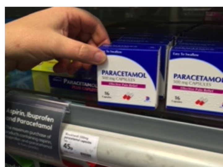
She was planning to move into a flat with her new boyfriend, but yesterday found out he was seeing someone else. Life seemed totally hopeless and she drank half a bottle of vodka and took thirty paracetamol tablets.

paracetamol is sold in boxes of only 16 in the UK to reduce the likelihood of overdose

She left the empty packets on the kitchen table – she admitted that she wanted her Mother to find them when she returned from the pub with Ken. They found her in her room, semi-conscious from the vodka, and rang for an ambulance.

In the emergency department she had blood taken, and the paracetamol level was high enough to need treatment. She was started on a drip containing acetylcysteine (Parvolex), about five hours after taking the overdose, and sent to our assessment ward.

December is always a popular month for deliberate self-harm. It might be the “dark days before Christmas”, with insufficient sunshine to activate the pineal gland. It also seems that Christmas puts a lot of tension on families, and makes some people very miserable, rather than happy. My Mother-in-Law remembers Christmas as a child:

“the smell of sprouts and the sound of weeping from the kitchen”

We have medical students on the ward, who have all recently spent time in primary care. I ask

“Do you see a lot of depression in GPs surgeries?”

They smile as they recount the very large numbers of patients who attend their family doctors because they are unhappy and seem unable to cope with their lives.

We look after a lot of people who have taken a deliberate overdose of drugs. Many are young and female, and when we go to see them in the morning, they are often hiding under the bedclothes – reluctant to talk. Sarah was not keen to talk to begin with, but soon told us the whole story. She said she felt really stupid now, and felt she had wasted everyone’s time. She was glad she had not managed to kill herself. We told her that she would soon be seen by our psychiatry colleagues. They sent her home later that day, after we had checked her blood results and were sure she had not done any serious damage to her liver. She has an outpatient appointment with the psychiatric team next week.

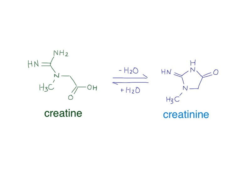
Overdose of paracetamol, or acetaminophen as it is called in the US, is the commonest reason for acute liver failure needing liver transplant in the UK and US in young people. Why is it so toxic in overdose?

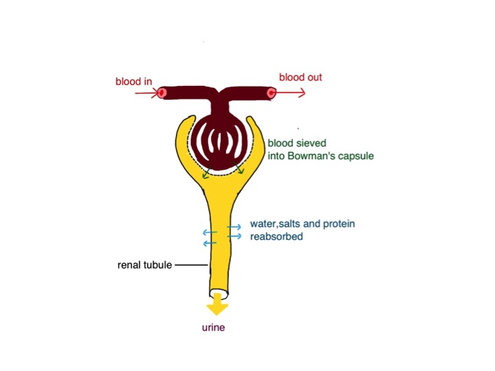
Paracetamol is metabolised by our liver. Normally, about 90% is conjugated with glucuronic acid or sulphate. Glucuronic acid is like glucose, with an extra COOH attached.

liver metabolic pathways for paracetamol
liver metabolic pathways for paracetamol – Fvasconcellos  wikicommons

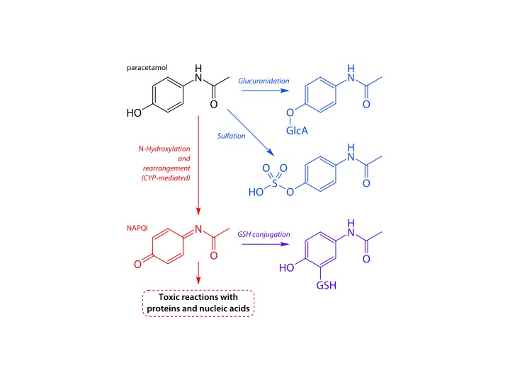
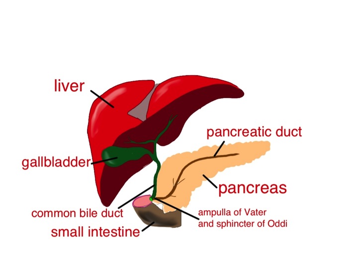
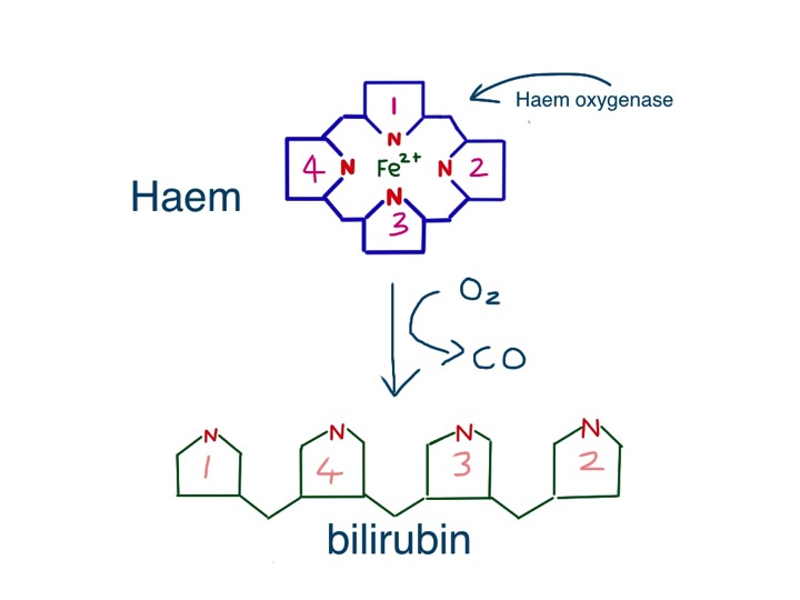
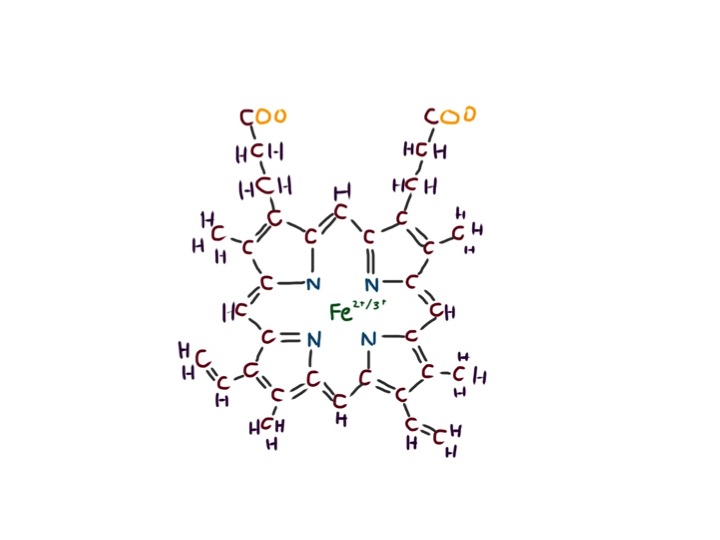
Once conjugated paracetamol is much more soluble in water, and is excreted in the kidneys. But about 10% is metabolised by liver cytochrome enzymes. They are called cytochromes because they make the liver cells coloured – a brownish red colour. Cytochromes enzymes contain haem, but mostly with the iron in the Fe3+ form. This makes them brownish rather than the bright red in haemoglobin and myoglobin, where the iron is in the Fe2+ oxidation state. That is why liver is a different colour from ordinary meat. These cytochrome enzymes are also known as p450 enzymes. This is because if a pure solution of the enzyme in a test tube is bubbled with carbon monoxide, the enzyme absorbs light with a peak at 450nm – purplish blue. Absorbing blue light means that it looks reddish – the colour which is not absorbed. There are a whole range of cytochrome p450 enzymes in the liver – to deal with the vast number of chemicals which our bodies need to deal with. Their job is to oxidise the variety of chemicals in our diet and render them soluble and make it easier for them to be excreted by our kidneys.

The small proportion of paracetamol which is oxidised by p450 enzymes is turned into a toxic chemical – NApQI – this stands for N-acetyl-p-quinone-imine. This molecule is only very slightly different from the parent paracetamol molecule, but contains a C=O group on the benzene ring, making it a quinone. Quinones are often reactive chemicals.  They are used industrially to generate hydrogen peroxide. NApQI will rapidly damage the liver cells that make it from paracetamol. But this damage does not happen when we take the normal dose. Fortunately all of the NApQI is rapidly detoxified by reaction with glutathione, a substance normally present in the liver cells, to produce a non-toxic conjugate which is excreted in the urine.

The problem comes when paracetamol is taken in overdose. The glucuronic acid and sulphate conjugation pathways are overwhelmed, and more of the paracetamol is turned into NApQI by cytochrome p450 enzymes. After a short time the glutathione runs out and NApQI cannot be detoxified and causes liver damage. The acetylcysteine we gave to Sarah as soon as she came in is used by liver cells to replenish glutathione and prevent any more liver damage. We give a lot of acetylcysteine – about 25 grammes in total – about an ounce, over a period of 21 hours. After this time all of the paracetamol should be eliminated as non-toxic substances. Chemicals that cause induction of p450 enzymes, such as alcohol and anticonvulsant drugs, can make paracetamol toxicity more likely. Malnutrition, which can deplete glutathione reserves, can also increase the risk of liver damage.


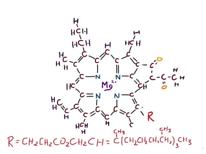
The second question I will ask, but not give a satisfactory answer to, is why does it take a long time for antidepressant drugs like sertraline to work? This drug, like Prozac, or fluoxetine, is thought to increase the amount of serotonin available in the brain. Serotonin is a small molecule, made from the amino acid tryptophan. There is a lot of tryptophan in certain foods such as soyabeans and cheese, but smaller amounts in all protein sources. Sertraline is an SSRI – a selective serotonin reuptake inhibitor. Serotonin is released from one nerve and travels across a synapse to cause an effect of the next nerve. Following release serotonin is taken back up into the cell that released it, causing the effect to terminate. Blockade of this uptake will mean that there is more serotonin available to have its effect. Drugs such as ecstasy (MDMA) are thought to have their euphoric effect by increasing serotonin levels, but they work more or less straight away.

I think we have an awful lot to learn about how the brain works. In fact I’m sure that we have very little idea. We don’t even seem to understand the basic operating system.

The information for this red apple is coded in the silicon chips of your computer as 0s and 1s, in a particular sequence to allow the screen to form this image. How does it work in your brain?

If I ask you to close your eyes and think of an apple, a nice red one, that is easy to do. But what is that thought? The thought – the image of an apple in your mind is certainly a thing – a physical thing. Although you think you are only imagining the picture of the apple – the picture must be there – somewhere. The thought must be made of something – even if it is a perturbation of electrical charge or change in chemicals. And it is certainly in your brain. But if I ask you exactly what the thought is made of, and where in your brain it is, we have no idea. No idea at all. Is the thought of an apple in the shape of an apple? – probably not, but of course it might be.

I have an analogy for how much we understand how the brain works. Let us imagine that you find an Amazonian tribe which has had very little contact with industrialised  society. You take them a plasma-screen television, a large solar panel to recharge the batteries, and a satellite dish. You put them together in a clearing in the forest, charge up the battery and turn the television on. Adjustment of the satellite dish results in the face of Simon Cowell presenting the X-Factor appearing on the television.

Simon Cowell photo courtesy Alison Martin of SimonCowellOnline.com.

The tribespeople are intrigued. You leave them with the whole kit. They start experimenting with it – they realise that moving the satellite dish changes the clarity of the image, they realise that they can change the picture or increase the sound volume by pressing various buttons. One enterprising young man has even unscrewed the back of the television and discovered that some components are removable, and taking them out does all sorts of interesting things to the picture and sound. They feel that after a few months they have a pretty good idea about how the television works. That is about the level of understanding we have of how our brains work – we can give people chemicals which change conscious level and behaviour, we can operate and stimulate certain parts of the brain to see what happens, and can use functional MRI to see what part lights up when we do, or think about certain things. But we don’t know how it works. Not a clue.

Now for the food link – grapefruit.Slide6 We have known for some time that drinking grapefruit juice can cause potentially serious interactions with certain drugs which are metabolized by liver p450 enzymes. This was discovered when researchers, who were looking for an effect of alcohol on the blood pressure drug felodipine, mixed the alcohol with grapefruit juice to disguise the taste. They found that the grapefruit juice, rather than the alcohol, had a huge effect on the rate at which felodipine was metabolized by the liver. This is because it contains a substance which binds to and inhibits certain cytochrome p450 enzymes.  Grapefruit also affects the metabolism of many other drugs, such as statins and oral contraceptives – for more information see: http://www.healthcentral.com/peoplespharmacy/pp_guides/pdf/gfruit02.pdf

gallstones and mayonnaise

A few weeks ago we admitted Sylvia. She had been feeling unwell for a some days, with fever and episodes of uncontrollable shaking. What really alarmed her was when she turned yellow, and then developed abdominal pain and vomited a couple of times. Sylvia is forty eight, and had always been healthy. She rowed with the local club twice a week and kept her weight under control. For the past year or so she noticed that she got pains in her abdomen after eating fatty foods such as fish and chips, and now avoided foods like that. It was her son who noticed she was yellow – he then phoned Sylvia’s GP. The duty doctor came and confirmed that she was indeed jaundiced, feverish and unwell, and persuaded her that she needed to go to hospital.

these people look jaundiced - in fact they are not - the whites of their eyes are white - this image is copyrighted but Wiki thinks its ok to use it so I'm hoping its ok for me to do so
these people look jaundiced – in fact they are not – the whites of their eyes are white – this image is copyrighted but Wiki thinks its ok to use it so I’m hoping its ok for me to do so

We did all the usual things for someone with suspected biliary sepsis. Took blood cultures and routine blood tests. These tests confirmed she had elevated bilirubin and transaminases, increased alkaline phosphatase, as well as a high neutrophil count and CRP.  We gave her intravenous fluids, anti-emetics and antibiotics. We were able to get an ultrasound within a couple of hours. Ultrasound is really good at looking at the liver and biliary system in jaundiced patients – better than CT.

The ultrasound showed that Sylvia had a gallbladder full of gallstones and her common bile duct was dilated. It seemed likely that she had a stone blocking the flow of bile from the liver to the duodenum, and that the stagnant bile had become infected with bacteria causing ascending cholangitis and liver inflammation.

bile is made in the liver - it is stored in the gallbladder and squirted into the small intestine after a fatty meal, along with pancreatic secretions - Sylvia's gallstone was impacted in the sphincter of Oddi
bile is made in the liver – it is stored in the gallbladder and squirted into the small intestine after a fatty meal, along with pancreatic secretions – Sylvia’s gallstone was impacted in the sphincter of Oddi

The questions I want to answer this week are:

Why did Sylvia turn yellow?

What are gallstones made of and why do they form?

Why is there an increase in alkaline phosphatase in the bloodstream when the biliary system is obstructed?

The last of those questions is the hardest, but I will make some conjectures, and  hope that others who know more about it will correct me.

Jaundice is fairly straightforward. It is caused by the accumulation of bilirubin, which is a greenish-yellow colour. Bilirubin is made from haem, the central working part of myoglobin, but most bilirubin comes from the breakdown of haem in the haemoglobin from red blood cells.

So why is haemoglobin red? It is not only – or even mainly – the iron that makes haem red, but the porphorin ring that surrounds the iron. This large ring is made of four smaller pyrrole rings.

Pyrroles are often coloured – the dye indigo has two pyrrole rings and red acrylic paint often contains the dye pyrrole-red. Melanin, the pigment that gives skin and hair its colour is brown or black or red because it has a related structure – indole – a pyrrole stuck to a benzene ring. Without this pyrrole ring we would be like albinos, but without pink skin and eyes.  As we shall see, we would also produce beige poo and colourless urine.

this is a pyrrole - all pyrroles are highly coloured, and are in most of the things which make people the colour they are
this is a pyrrole – all pyrroles are highly coloured, and are in most of the things which make people the colour they are

Red blood cells last on average 120 days before they wear out and have to be recycled.  As we have five litres of blood and in each litre there is at least 120 grammes of haemoglobin, it means we have to make, and break down, five grammes a day.  About a teaspoonful.  This all happens inside macrophages in the spleen and liver, as they try to salvage useful parts of the red cells to make new things.

Macrophages are similar to neutrophils but slightly smarter.  Some are good at host defence: they come to the scene after the neutrophils have done their job, do the forensics, and find out what the germs are made of. They then tell the lymphocytes how to make antibodies to protect us in the future.

Other macrophages are good at clearing up the mess, breaking down dead tissue and making things neat and clean.  The spleen and liver macrophages that deal with old red cells are the latter kind. When they take red cells apart, they separate haem from globin, and then take the iron out of the haem.  Next, the porphorin ring is broken to form bilirubin – its molecule of linked pyrrole rings looks like four beach huts in a row.

haem is broken down by the enzyme haem oxygenase to form bilirubin

The bilirubin is then thrown away (I don’t know why it can’t be re-used).  It attaches to albumin and becomes water soluble after conjugating in the liver with a sugary molecule called glucuronate.  The conjugated bilirubin is then transported into the biliary system, stored in the gallbladder, and squirted into the duodenum when we eat fatty food.  In the bowel, it is converted to urobilinogen, which makes poo brown.  Some is reabsorbed and excreted in urine, which makes urine yellow.Slide2

Sylvia had noticed that her poo had changed colour – it had become putty-coloured – and her urine had become much darker. That is because conjugated bilirubin was not getting into her intestines and instead some had leaked into her bloodstream and was appearing in her urine. The whites of her eyes and skin had become yellow because of the very high level of conjugated bilirubin in her blood.

Now to the gallstones.  In Western countries they are most commonly made of cholesterol.  I have already talked about the importance of cholesterol in keeping cell membranes rigid and non-leaky.  Cholesterol is made in large amounts by the liver and transported to other cells gift-wrapped as LDL cholesterol.  It is also used to make bile salts.  It only needs a minor modification of the basic chemical structure of cholesterol to make bile salts – the main component in bile which makes it work – the main function of bile salts is to help us digest dietary fat.

structure of cholesterol

Bile salts are detergents.  Just like washing-up liquid, they emulsify fats, breaking large fat globules into smaller micelles which do not stick to each other, or anything else. Cholesterol is essentially very insoluble in water, or hydrophobic.  When an organic acid group is added – to make bile salts – it gains a hydrophilic or water-attracting group. This is just like soap – a long lipid chain with an organic acid group at the end.  The hydrophobic lipid part is embedded in the tiny fat globule and the hydrophilic groups stick outside in the watery medium of the intestinal contents.

bile acids are a minor modification of cholesterol - hydrophilic OH  and COOH  groups are added to make it into a detergent
bile acids are a minor modification of cholesterol – hydrophilic OH and COOH groups are added to make it into a detergent

As well as bile salts, the liver secretes phospholipids into bile – principally phosphatidylcholine. This, with cholesterol, is the main stuff cell membranes are made of.  It is also an emulsifying agent, and is used extensively in the food industry to keep fats in suspension.

So, when we eat fat, the small intestine detects it and releases the hormone cholecystokinin (CCK).  This causes the gallbladder to contract to send bile into the duodenum, and also makes the pancreas release enzymes, including lipase.  CCK also has some very interesting effects on our brains, making us feel less hungry, and curiously, opposing the effects of opiate drugs.  The bile and pancreatic secretions are both delivered into the duodenum through the same tube – see diagram. The detergent bile salts, emulsifying phospholipid and pancreatic lipase are designed to work together to digest fat.

the hydrophilic parts of cholesterol are all on one side of the molecule - this allow it to act as a detergent - it works well to disperse fat into small globules so that pancreatic lipase can work on it and break it down to fatty acids and monoglyceride
the hydrophilic parts of cholesterol are all on one side of the molecule – this allow it to act as a detergent – it works well to disperse fat into small globules so that pancreatic lipase can work on it and break it down to fatty acids and monoglyceride

The bile breaks it up into tiny globules and the lipase breaks triglyceride into fatty acids and monoglyceride. These are transported across the intestinal mucosal cell membrane and then the triglyceride is put back together again. Seems a daft system, but it clearly works. The triglyceride is taken away from the intestine not in the portal blood, like most other substances absorbed by the gut, but in the lymphatic system in the form of chylomicrons – small fatty globules covered by a layer of phospholipid. The chylomicrons travel up the lymphatic vessels to emerge into the circulation just below our left clavicle. This means that it avoids passing straight away through the liver. If blood is taken soon after a fatty meal and centrifuged, it will have a milky appearance because of the large amount of chylomicrons.

So why do gallstones form? As well as bile salts, unmodified cholesterol is also secreted into bile. It is only kept soluble by the bile salts and phospholipid – the ratio of cholesterol to bile salts and phospholipid is therefore important. When it goes wrong, cholesterol precipitates out and forms solid stones like those that caused all Sylvia’s recent problems – a bit like when I try to make mayonnaise and it curdles – the fat becomes un-emulsified and separates out.

In some parts of the world, gallstones are made from bile pigment – bilirubin. This can be due to increased red blood cell breakdown resulting from abnormalities such as sickle cell disease, but it’s also common in East Asia, for reasons which are not clear.  A good, comprehensive analysis of why gallstones of all types form can be found at:

http://gastro.ucsd.edu/fellowship/materials/Documents/Gallstones/pathogenesis%20gallstones.pdf

Now the alkaline phosphatase. Bile is alkaline.  Alkaline phosphatase is an enzyme that removes phosphate groups in alkaline conditions.  When bile flow is blocked – by stones, as in Sylvia’s case, or for any other reason – the liver makes much more alkaline phosphatase, some of which appears in the blood.  Bones use a similar alkaline phosphatase to rearrange phosphate groups to make hydroxyapatite – the hard stuff bone is made from.  Most of the alkaline phosphatase normally in our blood is the bone sort.

There is plenty of information about whether alkaline phosphatase is from bone or liver, but very little I can find that suggests why the liver should make this enzyme when the biliary system is blocked.  I think a clue here is that neutrophils also have alkaline phosphatase in their granules, which help kill bacteria.  This is in addition to the myeloperoxidase and esterase mentioned in earlier posts.  Why should a phosphatase be damaging to bacteria?  The answer may well involve techoic acid, an important reinforcing molecule in some gram-positive bacteria.

cartoon of gram positive bacterial cell wall - techoic acid is thought to inhibit the action of lysozyme and protect the peptidoglycan from being broken down - techoic acid contains phosphate groups which may be removed by alkaline phosphatase
cartoon of gram positive bacterial cell wall – techoic acid is thought to inhibit the action of lysozyme and protect the peptidoglycan from being broken down – techoic acid contains phosphate groups which may be removed by alkaline phosphatase

In these microbes, techoic acid strengthens the peptidoglycan cell wall and inhibits the action of lysozyme, yet another enzyme made by neutrophils.  Lysozyme is designed to break some of the sugar-sugar bonds in peptidoglycan – so getting rid of the techoic acid would surely be helpful.  We know that alkaline phosphatase is effective in breaking up techoic acid – see, for instance:

http://jb.asm.org/content/102/3/747.short

Maybe the liver is producing this anti-bacterial enzyme to help prevent infection when the flow of bile slows down.  Unfortunately, this won’t work with gram negative organisms such as E.coli because they don’t make techoic acid – and it was a gram negative bacterium we grew from Sylvia’s blood culture – the germ that was causing her ascending cholangitis.

Soon after she came in, Sylvia had an ERCP – an endoscopic retrograde cholangio-pancreatoscopy.  The endoscopist managed to remove the impacted gallstone from her ampulla of Vater, and she quickly recovered.  A week later she had her gallbladder removed, including the stones, and now is quite well – she can even eat fish and chips.

Now to the food link: mayonnaise.

French mayonnaise - made with olive oil and mustard
French mayonnaise – made with olive oil and mustard

This is an emulsion of lipid, such as olive oil, in an aqueous (watery) medium – vinegar or lemon juice.  The emulsifying agent is raw egg yolk, which – just like bile – contains lots of phospholipid – phophatidylcholine and is also rich in cholesterol.  In France, mustard is also added.  Mustard seed contains mucilage, a gooey stuff that some plants produce made of sugar polymers. (Thanks again to Harold McGee). These also act as emulsifiers and further thicken the mayonnaise, and give it a better taste. When I make cheese sauce, I use a sugar polymer (flour) to achieve a stable emulsion between butter and milk – no doubt the phospholipid in the milk helps too.  It seems likely that in the small intestine, the mucus from the stomach, a similar gloopy sugar polymer, probably has a similar effect to mustard seed mucilage to help with fat emulsification.

Broken arm and salami

The NHS is having a bit of a crisis in the UK, struggling to look after an increasingly frail, elderly population. This week we admitted Agnes. When I saw her name in the casenotes I knew she was going to be elderly. Some names popular in the 1920s have become fashionable again, such as Daisy, Sophie and Amy. Not so for Agnes. She is 93 and was just about managing at home on her own. She has diet-controlled diabetes and recently had a hip replacement.

She slipped when getting out of the bath and fell on her outstretched hand, breaking her right wrist. The alarm she normally keeps around her neck was on the shelf over the bathroom sink. She did not have the strength to get up and nobody heard her calls for help. Agnes lay on the floor for more than a day without food or water.  Eventually her three-times-a-week carer called and found her and called the ambulance.

typical Colle's fracture of the wrist - from Wikimedia commons by
typical Colle’s fracture of the wrist – from Wikimedia commons by Ashish j28

She was already a bit happier when she arrived in the emergency deparment after she had been given some fluids and pain relief by the ambulance crew. She was very sore down one side of her body with obvious large bruises over her buttocks, thighs and arms. The emergency department doctors and nurses had fixed her wrist and put it in a plaster.

Her blood results were worrying. Her creatinine level was raised to over double the normal value for her age, and much higher than it had been after her hip operation 3 months ago. Her creatine kinase level was over 20,000, the normal being below 150.

We were worried because muscle damage can cause severe kidney damage. We knew she had a lot of muscle damage because her creatine kinase level was so raised. Creatine kinase is an enzyme which is vital for normal muscle function. Kinase means an enzyme which adds a phosphate group to creatine. Creatine and creatine phosphate are small molecules which are found in large amounts in muscle – what does creatine do?

Muscles need energy to work. This is supplied by mitochondria which use glucose and other energy sources such as fats and protein. If you throw sugar or fat on a fire it will burn rapidly and provide heat. Mitochondria “burn” energy sources by eventually combining them with oxygen in a much more controlled way. Instead of producing heat, the energy is converted into a chemical bond. Adenosine di-phosphate (ADP) is converted into ATP – adenosine triphosphate. Addition of this extra phosphate is rather like pulling the bowstring on a crossbow. The energy is ready to use to fire the bolt when needed.

The energy stored in ATP is in the chemical bond of the third phosphate group. This energy-containing ATP can then be used by the cell to make muscles contract or perform a whole host of housekeeping functions such as making cholesterol (see previous post).  Muscle needs lots of ATP, so muscle cells have lots of mitochondria. The problem is that ATP does not store well. That’s where creatine phosphate comes in handy. It stores well and can rapidly regenerate ATP from ADP.

creatine and creatinine are small molecules which are in chemical equilibrium

Creatine phosphate is sold in health food shops. There is some evidence eating it can improve exercise performance by increasing creatine in muscles. Creatine is spontaneously converted to a similar molecule, creatinine. This is produced in similar amounts in all of us every day, depending how much muscle we have got, and excreted unchanged by the kidneys.

So why is the small molecule creatinine excreted in kidneys, but molecules we want to keep are not? Kidneys work in a strange way. When we want to get rid of unwanted rubbish in our houses we find the things we don’t want and put them in the bin and take them out to be collected by the waste disposal team. Kidneys do it differently. They do the equivalent of taking all the contents of our house (not large things like furniture) and putting them in the garden. Then they find all the things they want to keep – such as water, sodium, potassium, small proteins and hydrogen ions, and take them back in the house.

pigs kidney
pigs kidney

Before she fell over, Agnes’s kidneys were filtering about 80mls/min through her kidneys. We can calculate this from knowing her age, sex and creatinine level using a modification of a really useful formula devised by a Canadian chest doctor Donald Cockcroft.  You can see a picture of him here:

http://www.medicine.usask.ca/medicine/divisions/respirology/faculty/donald-w.-cockcroft.html

His paper has become a citation classic:

Click to access A1992JX46100001.pdf

Doctors talk about kidneys filtering fluid in the kidney.  What they really mean is that blood is sieved, like when you put boiled bones, onions and carrots through a sieve to make stock.  The kidney is not one big sieve, but instead it does the sieving using about a million mini-sieves. This happens in tiny structures called glomeruli where the pressure of arterial blood forces water and small molecules through the sieve into the Bowman’s capsule (see diagram).

cartoon of a glomerulus and renal tubule - modified from Wiki commons madhero 88
cartoon of a glomerulus and renal tubule – modified from Wiki commons madhero 88

The sieve has very small holes which do not allow red cells, white cells, platelets or most protein to pass through (that’s the furniture).

Eighty millilitre per minute is a lot of fluid. Nearly 5 litres every hour. Clearly we can’t afford to lose that volume of fluid so all the water is reabsorbed in the renal tubules (see diagram). The sieved fluid goes along a long tube lined with cells whose job it is to pull the water and vital salts back into our body. It might appear to be a daft system but (a) it works and (b) has the advantage that any new or foreign substance will be eliminated by the kidney if it can pass through the glomerular sieve. It means we don’t have to be able to recognise a chemical to dispose of it – The kidney tubular cells just say – “I need that – we’ll bring that back in”. Our house could do with a system like that.

So why have Agnes’s kidneys stopped working? The problem is that she has damaged a lot of muscles by lying on the floor and the protein myoglobin has leaked out of the muscles into her bloodstream and stuck in her kidney tubules where it causes damage. Some muscle has lots of myoglobin – it is what makes meat red.

When people talk about a bloody steak, it’s not blood which makes the meat red and drip red fluid, it is myoglobin. Myoglobin is like haemoglobin.

Haem is the key working part of myoglobin and haemoglobin. The iron is trapped safely in the middle

Its function is to help drag oxygen out of the bloodstream, where it is attached to the haemoglobin of red cells, and deliver it to the mitochondria. It is very similar chemically to haemoglobin- it has a haem group attached to a protein (heme for US readers). Haem is very useful, it is a chemical structure containing iron which keeps this reactive element under control. Like a rotweiller on a leash. Haemoglobin and myoglobin are both bright red, not only due to the iron, but mainly due to the porphyrin rings which make up haem. For iron to work in haemoglobin and myoglobin it must be in the reduced, ferrous or Fe2+ form. It wants to be oxidised to the Fe3+. Myoglobin with iron in the Fe3+ is known as metmyoglobin, but is kept reduced Fe2+ in red cells and muscle cells by enzymes especially designed for that purpose (metmyoglobin reductase).

rump steak goes brown on the surface due to conversion of myoglobin into metmyoglobin

Myoglobin in dead muscle (meat) rapidly becomes oxidised to metmyoglobin. This Fe3+  form of myoglobin is brown. That is why when you buy steak it is brown on the surface but red in the middle where there is not enough oxygen to form metmyoglobin. Heat rapidly causes oxidation, which is why steaks go brown when they are cooked. Not all muscle contains myoglobin. Chicken breast is pale because it has little myoglobin. Genetic knock-out techniques have bred mice which have no myoglobin in their muscles and amazingly they seem to be relatively healthy – see

http://www.nature.com/nature/journal/v395/n6705/abs/395905a0.html

Myoglobin is a very small compared with most proteins. It is smaller than the holes in the sieves in the kidney. When muscle is damaged myoglobin leaks into the circulation. Small amounts are bound by another larger protein called haptoglobin. When all the haptoglobin is used up free myoglobin goes through the kidney sieve. Other small proteins do this and are rescued back into the body by the renal tubular cells. The problem is that the iron in myoglobin, when it escapes from muscle cells is rapidly oxidised to metmyoglobin. The Fe3+ then displays its unrestrained rotweiller tendencies – it starts to cause damage. Elemental iron engages in Fenton reactions which generate free radicals.

rust is reddish brown because it is iron in the form of Fe3+

What biomedical scientists call a free radical, chemists call a radical. It is a compound with an unpaired electron, and therefore usually very chemically reactive. Iron is a transition metal, which means that it is very happy to gain or lose one electron at a time. Iron in the form of Fe3+ lodged in the kidney tubules causes a lot of free-radical damage making the tubule cells swell up and obstruct the flow of urinary filtrate. More details in this free full-text paper by Kevin Moore:

http://www.ncbi.nlm.nih.gov/pubmed/9822635

Because Hilda’s kidney tubules contain a lot of metmyoglobin, her urine is a dirty brown colour – an important clue that muscle damage, also known as rhabdomyolysis, may be part of the problem.

We treated her with intravenous fluids and her kidneys recovered over the next 4 days. She went to stay with her 95 year old sister Phyllis because she could not manage with only one arm working. She was not looking forward to it – Phyllis still treated Agnes like a irritating little sister after all these years.

I am writing this on a train travelling through Devon in the UK. The soil is a wonderful red-brown colour, covered with bright green trees and grass.

seaside cliffs in Devon are red because of lots of iron deposited as Fe3+

The soil is red because it has lots of iron in it. Much of the iron in our soil was deposited there in the “great oxygenation event” (not, as you might think at the 02 arena).  This happened about 2.5 billion years ago. Before this the atmosphere contained no oxygen –it was mainly nitrogen, methane and carbon dioxide. The primitive organisms – anaerobic bacteria, are killed by oxygen. Then came along cyanobacteria which contain chlorophyll. Bright green chlorophyll is a remarkable molecule which is able to convert carbon dioxide and water to the very useful molecule glucose. A side product of this chemistry is the formation of oxygen. This oxygen oxidised all the iron in the oceans from green Fe2+ to the rusty-coloured Fe3+ which is less soluble and precipitated to make our soil red. When all the iron was oxidised, oxygen appeared for the first time in our atmosphere, and is still being maintained by plants containing chlorophyll.

England would look very different without chlorophyll

The structure of chlorophyll is really quite similar to haem.

chlorophyll has a very similar structure to haem, except it has magnesium in the middle instead of iron

It has a porphyrin ring, but incorporates a magnesium atom instead of iron. If you boil your vegetables for too long the magnesium falls out into the cooking water and your greens turn muddy yellowish-gray. Victorian cooks would put a copper penny in their cooking water – copper replaces the magnesium and keeps the greens green. Don’t try this at home, but do read Harold McGee’s book “On food and cooking”. It tells you everything you ever wanted to know about the science of cooking and is totally readable if are interested in how the world works.

Finally the food link – salami. Salami is usually pink.

very pink salami
very pink salami

This is because it is preserved with nitrite. Salami can be made with all sorts of meat, including donkey. Without nitrite the meat would turn brown when minced due to methaemoglobin formation as explained above. Nitrite reacts with acids in the meat to form nitric oxide which combines strongly with myoglobin to form nitrosomyoglobin. Nitrosomyoglobin is bright pink, which is why salami, corned beef, bacon, ham are that colour and do not discolour with storage.

new edition of harold mcgee's book - updated and more comprehensive, but more of a pain to carry around
new edition of harold mcgee’s book – updated and more comprehensive, but more of a pain to carry around than the first edition

Nitric oxide is also a useful stuff for killing nasty anaerobic bacteria which can cause serious disease such as botulism. Clostridium botulinum is a germ which forms spores that will hatch and grow in the anaerobic (oxygen poor) atmosphere in corned beef cans, and make botulinum toxin – also known as botox. Botox is arguably harmless when injected in tiny amounts into the face of rich, vain women, but very bad when swallowed in large amounts to cause paralysis of all our body muscles leading in death from respiratory failure. The main reason nitrite is still allowed in preserved meat products is that it can prevent botulism.

I learned today that Hugh de Wardener died on September 29th age 97. You will remember that I wrote about him recently in the article vodka and sweetbreads. There is an obituary at http://announcements.thetimes.co.uk/obituaries/timesonline-uk/obituary.aspx?n=hugh-edward-de-wardener&pid=167291852#fbLoggedOut