Category Archives: Uncategorized

falling over and liquorice

We admit a lot of elderly patients who have fallen over. There are a whole range of problems which cause falls, one of which is postural, or orthostatic hypotension.  Emily is 78 and lives alone in a bungalow. Her husband died three years ago, but her daughter calls in to help most days. She was fairly well until she went to her family doctor’s surgery last week and had her blood pressure measured.  The result was not good – 186/88mmHg.

She first developed high blood pressure when she was in her fifties, and it had crept up since. She was already taking ramipril (an ACE inhibitor) and indapamide (a thiazide diuretic). She had tried amlodipine, but that gave her swollen ankles, which she hated. Her GP decided to try her on doxazosin. This is an alpha-blocker that lowers blood pressure by opening up small arteries. The morning of the day of admission she got up and took her tablets. Then she had a bath, but when she got out felt awful, and faint, and fell to the floor. She lay on the floor for a few minutes and tried to get up, but felt very dizzy and had to lie down again. She phoned her next-door neighbour on her mobile phone. The neighbour came round and helped Emily to her feet, but she went wobbly and could not stand, ending up on the floor again. Her neighbour became really worried and called for the ambulance. When the ambulance paramedics arrived they found that when they tried to stand Emily up, her blood pressure dropped from 160mmHg systolic to 90mmHg. They brought her in to see us.

There is a popular notion that all antihypertensive drugs cause postural hypotension, but this is not the case. As we will see, beta-blockers can even be used to treat this condition.

What is happening with the circulation to cause Emily’s blood pressure to drop when she stands up?

standard clinical rubber glove

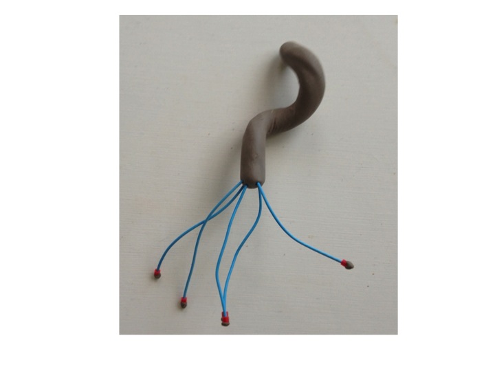
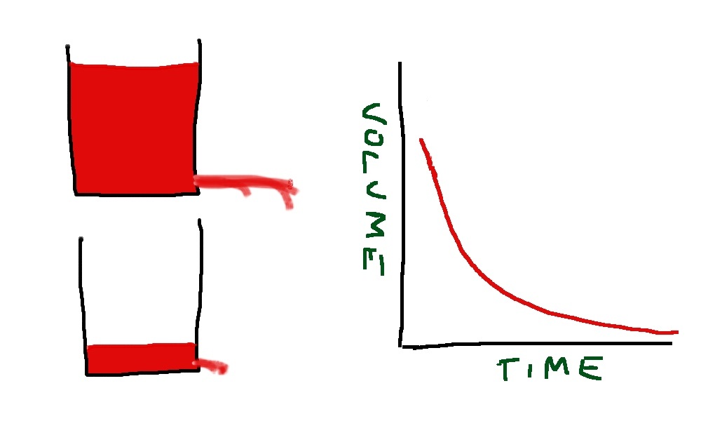
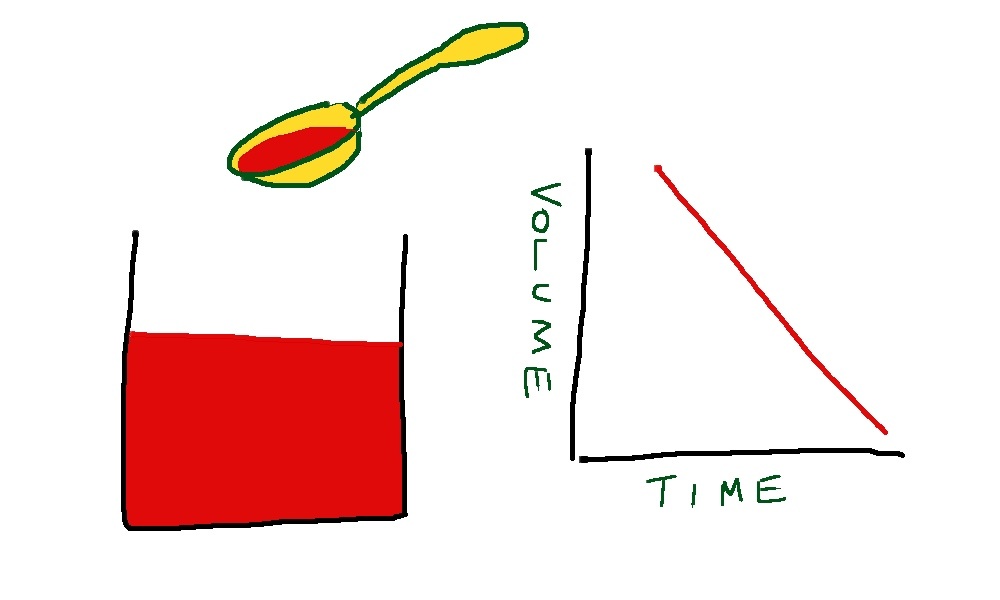
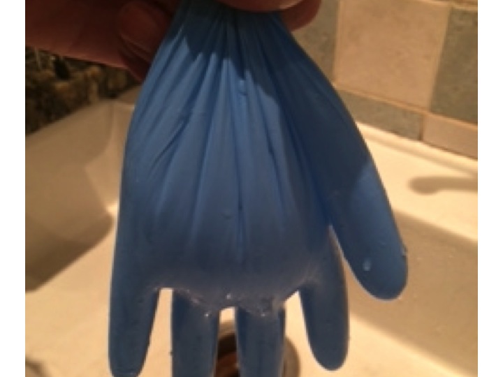
The whole explanation is made much easier with a standard hospital rubber glove (the stretchier the better) and a sink, and a supply of water. First we fill the glove up with the right amount of water.

glove about half filled with water - laid flat - notice the thumb is filled with water - this represents cardiac filling pressure
glove about half filled with water – laid flat – notice the thumb is filled with water – this represents cardiac filling pressure

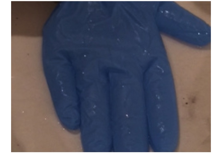
The water in the glove represents blood in the venous side of the circulation. The arterial side is much lower volume, and is very little affected by changes in pressure due to posture.

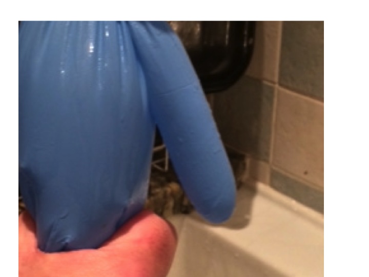
When we stand up the amount of fluid in the veins of the legs and abdomen increases, due to increase in hydrostatic pressure stretching the rubbery material. Veins are thin-walled and rubbery, and will naturally distend if the pressure inside is increased.  If this happens, the thumb empties, this represents the filling pressure to the heart. If cardiac filling pressure drops,  cardiac output drops and blood pressure falls.

no venous squeeze due to failure of the autonomic system, due to brain stem or peripheral problems means no cardiac filling pressure - the thumb is empty
no venous squeeze due to failure of the autonomic system, due to brain stem or peripheral sympathetic nerve action means no cardiac filling pressure – the thumb is empty

This will result in a fall in systemic arterial blood pressure – postural hypotension.

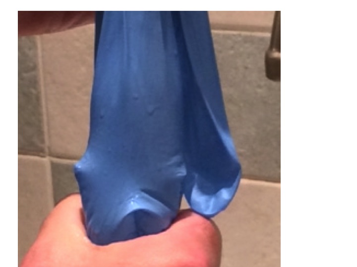
But this does not happen in healthy, young people. We have autonomic reflexes which cause constriction of veins before we stand up, so that filling pressure to the heart is maintained.  In the glove model this is represented by my hand squeezing the fingers and main hand of the glove.

in healthy individuals the autonomic nervous system provides a squeeze of abdominal and leg veins to maintain filling pressure of the heart - notice the thumb is now full of water
in healthy individuals the autonomic nervous system provides a squeeze of abdominal and leg veins to maintain filling pressure of the heart – notice the thumb is now full of water

There are two main causes for postural hypotension – failure of autonomic squeeze of the veins, and insufficient filling of the venous compartment.

Let’s take the first of these – not enough squeeze.  When we move from lying down to standing up, our brain knows what is about to happen. As we move our limbs the proprioceptors in our joints tell our brain what is about to happen and our conscious mind probably also has a role. Messages are sent to the control centre (Houston – prepare for lift-off) in the medulla. Sympathetic nerves send messages down to the veins to tell them to constrict. It is interesting that human intra-abdominal mesenteric veins are particularly richly supplied with sympathetic nerve endings – presumably because we are one of the few animals where standing is such a problem. In animals such as horses and dogs, moving from lying to standing does not involve a capacious venous network to be subjected to large pressure changes – their legs have very little venous blood and their abdomen and contents is on the same level as the heart.

It is also not surprising that if you take a healthy, young person and strap them to a tilt-table, and suddenly move them from horizontal to vertical, they will experience an impressive drop in blood pressure. If they are lying on a bed and stand up – blood pressure hardly changes because the brain-stem prepares us for the change in posture with messages to our abdominal and leg veins to constrict.

As we get older, everything starts to go wrong (see last week’s post). Autonomic reflexes become impaired. Patients with Parkinson’s disease are particularly prone to develop postural hypotension because of impaired brain stem autonomic reflexes – this used to be called Shy-Drager syndrome when I was young – it is now know as multi-system atrophy. Some drugs such as antidepressants and methydopa inhibit brain stem sympathetic output and predispose to this condition.

The sympathetic nerves end up on the outside (adventitia) of veins and release noradrenaline which causes constriction of venous smooth muscle by stimulating alpha receptors. Emily had been given an alpha-blocker to reduce constriction of her arterioles and thereby reduce blood pressure. In her it reduced the ability of veins to constrict when standing up by blocking venous alpha receptors.

Some patients have problems with the autonomic nerves. Diabetics and alcoholics are prone to develop autonomic neuropathy which may result in postural hypotension due to inability of veins to constrict.

Temperature also plays a part in venous constriction. Most of us have noticed occasionally when getting out of a hot bath or sauna feeling dizzy for a few seconds, we hold onto the edge of the bath then things improve. What has happened is that the hot bath water has caused venous dilatation and the normal reflexes have not been enough to maintain cardiac filling pressure. Cardiac output drops, baroreceptors panic (Houston, we have a problem). Houston responds quickly by sending a stronger sympathetic signal to the veins and to the heart to increase cardiac output and the problem is soon sorted. But of course preventing the problem is much better than reacting when it has happened.

The second main cause of postural hypotension is insufficient fluid in the circulation.

to represent fluid loss - some water is poured out of the glove
to represent fluid loss – some water is poured out of the glove

In Emily’s case this may have been partly due to the thiazide diuretic she was taking for her blood pressure. Patients on loop diuretics such as furosemide are even more prone to fluid depletion. Sepsis causes problems with a postural blood pressure drop because of fluid shifts out of the circulation into interstitial spaces, and because fever causes venous dilatation as in the hot bath above. Jonathan, who had a lobar pneumonia a few weeks ago, may well have collapsed in his GP’s surgery because of a postural blood pressure problem.

even with reduced volume - when lying flat the thumb is still filled with water and blood pressure is maintained
even with reduced volume – when lying flat the thumb is still filled with water and blood pressure is maintained

Blood loss from trauma or intestinal bleeding may often not result in blood pressure drop until the patient stands up – it is far easier to maintain filling pressure when lying down and not having to squeeze the veins hard.

with reduced volume, squeezing the legs and abdomen no longer maintains filling of the thumb - cardiac filling reduces and blood pressure drops
with reduced volume, squeezing the legs and abdomen no longer maintains filling of the thumb – cardiac filling reduces and blood pressure drops

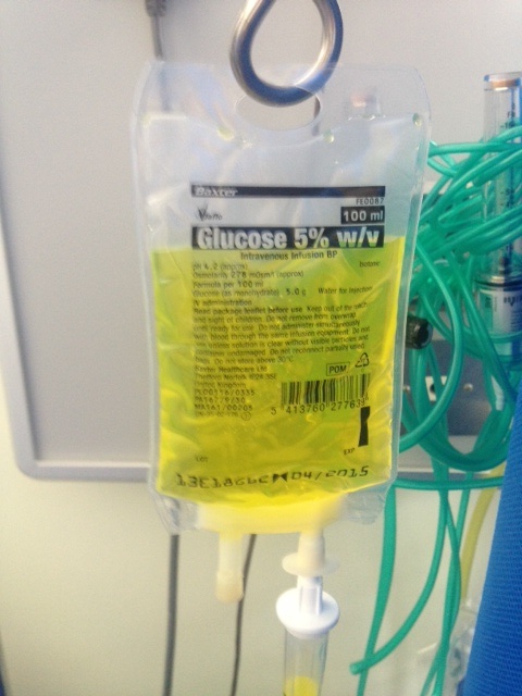
Sorting out Emily’s postural hypotension was straightforward. We gave her two litres of intravenous saline and stopped her doxazosin, and diuretics. We had a plan to start her on verapamil or diltiazem when she could stand up without her blood pressure dropping – which it did the next day.

Do ACE inhibitors/ARBs, beta blockers or calcium channel blockers cause postural hypotension?

The answer is to begin with, no and no.

ACE inhibitors can cause a postural drop when they are first started. This is because angiotensin does have an effect on venous constriction, but only transiently when it is increased or decreased. If angiotensin II is infused into a hand vein it will constrict, but only for a few hours at most. Similarly if angiotensin II is withdrawn, the vein will dilate, but only briefly. This means that it is prudent to warn patients that they may develop postural symptoms following the first dose – and it is a good idea to take it when lying down before going to bed. The effect of angiotensin II on arterioles is long-lasting, which is why these drugs are useful in treating arterial hypertension.

Beta blockers reduce blood pressure by mechanisms which are not completely understood, but is likely to be a combination of reduced cardiac output and inhibition of renin release from the kidney. There are beta2 receptors in arteries and veins, but these cause vascular dilatation. Beta blockers can help veins constrict, which is why they are sometimes used to treat postural hypotension. Having said that, if there is a postural drop in blood pressure, the reflex mechanisms set in train by baroreceptors involve increased sympathetic stimulation to the heart via beta1 receptors, increasing force and rate of contraction. Beta blockers will block this response and may therefore impair the recovery from an episode of postural hypotension, but they will not cause it.

Calcium channel blockers have an effect on arterioles, not on veins. Calcium channels are important in maintaining arterial tone, but not venous tone. Venous tone depends almost entirely on sympathetic stimulation. Therefore, at least in theory, drugs such as verapamil, diltiazem and amlodipine will not cause a postural drop. But of course if there is a postural drop, lower initial blood pressure caused by these agents may make the episode worse.

Sorting out Emily’s orthostatic hypotension was fairly straightforward. What about when it is caused by age-related impairment of brain stem function, or by irreversible peripheral autonomic neuropathy? It can be a real problem.

overfilling the glove means that even with no squeeze to the veins the thumb remains full of water when the glove is upright - overfilling the circulation with fludrocortisone can help prevent postural hypotension
overfilling the glove means that even with no squeeze to the veins the thumb remains full of water when the glove is upright – overfilling the circulation with fludrocortisone can help prevent postural hypotension

One approach is to use fludrocortisone. This is a synthetic analogue of aldosterone. Made from cholesterol (again!) this hormone is made in the adrenal cortex, in different cells which make cortisol from cholesterol. The function of aldosterone is to regulate how much salt and water is excreted from our kidneys. Aldosterone helps the reabsorption of more salt from the distal convoluted tubule and will therefore increase circulating volume of blood. This will have the opposite effect to diuretics and overfill the circulation and help keep filling pressure at heart level adequate on standing up. Another drug which is sometimes used is midodrine, an alpha receptor agonist, helping constrict veins and maintain cardiac filling when upright. The drug does not have a licence for this indication in the UK.

We have made a not very good video showing how postural hypotension works – my first attempt at youtube – thanks for your help Steph. We will try to make a better one soon and update this post. The video is at:

The food link this week was not obvious when I started, but of course it has to be liquorice.

liquorice has been popular for a long time - it is made from the root of the liquorice plant
liquorice has been popular for a long time – it is made from the root of the liquorice plant

Liquorice contains glycyrrhizin which has a chemical structure similar to steroids such as cholesterol and aldosterone (made from cholesterol – again!). It has long been known to have a mineralocorticoid effect. This is not because it acts directly in the kidney to stimulate aldosterone receptors. Instead it prevents the conversion of cortisol to cortisone. Cortisone, synthesized by the adrenal glands, is converted to cortisol in the kidney, then back to cortisone, cortisol has more mineralocorticosteroid effects than cortisone – full details are in the NEJM paper:

http://www.nejm.org/doi/full/10.1056/NEJM199110243251706

Eating too much liquorice can cause high blood pressure and low plasma potassium levels –  similar to primary hyperaldosteronism.

vomiting blood and yoghurt

Slide04 More alcohol problems this week. Peter came in vomiting blood yesterday afternoon. Vomiting blood is always a bad thing, but this time it was particularly bad. His long-suffering partner Rita came in with him to tell us what had happened. Peter, aged 69 was too drowsy and confused to tell the story himself. He was a retired barman and had always drunk too much alcohol. He recently went to see the liver doctors because his abdomen had filled up with fluid. They told him that he would die soon if he did not give up drinking. Rita said that he had cut down but was still drinking about 3 pints of strong cider every day. Yesterday lunchtime he was about to sit down to eat when he said he felt very sick. He staggered to the bathroom and promptly vomited what Rita estimated to be a pint of bright-red blood down the toilet pan. Rita called the ambulance. He vomited more blood on the way and by the time they arrived he was pale, sweaty and quite drowsy. She was really worried – she thought about what the liver doctor had said about Peter dying soon if he did not give up drinking – he had not given up.Slide05

When patients with liver disease vomit blood it always makes us worry about bleeding varices. Varices are large, distended veins which appear at the junction between the oesophagus (foodpipe) and stomach in people with liver cirrhosis. Cirrhosis often results from liver damage due to alcohol. Ethanol is metabolised to ethanal (acetaldehyde) and causes damage to liver cells as well as the pancreas (see vodka and sweetbreads below). The liver does have a remarkable capacity to regenerate.

Slide06

I’m not sure if the ancient Greeks knew about liver regeneration when they devised the myth about Prometheus. He made the mistake of giving fire to men, and as a punishment was chained to a rock for eternity. Every morning an eagle would fly down and peck out his liver. During the following day his liver would grow back again, to be pecked out again the following morning. He is still there.

ancient Greek vase showing Prometheus having his liver pecked out by eagle wikimedia common user Bibi Saint-pol
ancient Greek vase showing Prometheus having his liver pecked out by eagle –  wikimedia common user Bibi Saint-pol

Although liver does regenerate when damaged by alcohol, it does so to form nodules of liver tissue with bands of fibrosis in between the nodules. This disturbance of normal architecture impairs blood flow through the liver. As a result the pressure in the portal veins carrying blood from the stomach and intestines to the liver increases. Increase in portal venous pressure results in oesophageal varices. When they burst, rapid death from blood loss is common. Another result of increase in portal pressure is ascites – fluid accumulation in the abdominal cavity – the cause of Peter’s abdominal swelling.

all the blood coming from the stomach and small and large intestine goes into the portal venous system and through the liver to be processed - including removal of ammonia and small amines
all the blood coming from the stomach and small and large intestine goes into the portal venous system and through the liver to be processed – including removal of ammonia and small amines – from Grays anatomy 1918

So he was filled up with a blood transfusion, vitamins (see vodka blog below) and given terlipressin – a drug which constricts oesophageal varices and helps to stop bleeding. He was sent as an emergency to have an upper gastrointestinal endoscopy. In fact he did not have significant varices. He had a bleeding duodenal ulcer. The ulcer was cauterised and injected with adrenaline, a biopsy was taken from his duodenum, and he was sent to the admissions unit.  Part of the duodenal biopsy was put into a CLO test kit.

I talked previously about adrenaline causing muscle tremor and relaxation of bronchial smooth muscle by activating adrenergic beta receptors. It is released by the middle (medulla) of adrenal glands in response to severe stress. The reason Peter was so pale was probably more to do with release of adrenaline than blood loss. This hormone has many other actions to help us survive life-threatening situations. It will also act on beta receptors in muscle blood vessels to increase muscle blood flow – good to get away more quickly from the nasty tiger with dripping fangs that likes to eat humans.

Skin blood vessels have few beta receptors – here adrenaline acts on alpha receptors to cause reduction in blood flow. Similarly, in the lining of the duodenum, adrenaline, when injected by the endoscopist, causes blood vessels to constrict and help stop bleeding by acting on their alpha receptors.

When I ask students why adrenaline reduces skin blood flow they usually say it is to redirect blood to the central circulation where it is more needed. The skin only has about 1% of circulating blood. It is more likely that skin blood flow is reduced to limit blood loss when the tiger’s teeth finally sink into that tasty human flesh.

All this preamble is a good excuse to talk about urea. In our hospital normal blood urea levels are between 3.5 and 6 mmol/l. In the US doctors talk about blood urea nitrogen- the same stuff- normal levels are 20-30mg/l. When Peter visited the liver doctor last month his blood results showed that the concentration of urea in his blood was low – only 1.8 mmol/l (5mg/dl BUN). When he arrived in the ED it was elevated at 14 mmol/l (40mg/dl BUN). His haemoglobin was low at 90g/dl and clotting was deranged with an INR of 2.9. Why was his urea low before and now high? To understand this I need to talk about protein metabolism.

Most of us in the West eat lots of protein. More than we need. In the US and UK adults eat about 100grammes of protein/day, although we only need about 50. If we eat 100grammes of protein a day, we need to get rid of the same amount, unless we are growing, body-building or pregnant. Patients who are ill typically break down more protein than they take in – negative nitrogen balance.

I’ve used arguments about in/out balance for fluid in my previous post and will use it for energy in future posts. Not everything in humans can be understood in terms of in/out balance. For instance, with my 11 year-old son we input high grade educational material and all that comes out is poo and fart jokes.

So what happens to this 100grammes of protein? Protein is a polymer of amino acids. You can make useful plastic out of milk protein – see this youtube video:

http://www.youtube.com/watch?v=pIvAl4lu1uA

Casein also used to be used to make plastic items such as buttons. We now have cheaper and better plastics.

Protein that goes into our mouth is mashed up by our teeth and swallowed. The stomach has the first go at breaking up the protein polymer with the enzyme pepsin, secreted by chief cells in the glands of the stomach. It’s a bit tricky making an enzyme that breaks down protein, because all our cells are made of lots of proteins.  There is the obvious danger that the enzyme will destroy the cell that made it. So pepsin is made in an inactive form – pepsinogen that is only activated when it comes into contact with stomach acid. Then the pancreas has a go. It makes trypsin, carboxypeptidase and chymotrypsin which finish the job, to end up with amino acids. The pancreas has to be pretty careful too, making inactive enzymes which become active one secreted – see:

http://www.physiologymodels.info/digestion/proteins.htm

Amino acids are all of the general formula:

general formula for amino acid - the red box stuff can usually be turned into carbon dioxide and water - the blue box stuff is harder to get rid of
general formula for amino acid – the red box stuff can usually be turned into carbon dioxide and water – the blue box stuff is harder to get rid of

The R is mainly made of carbon, hydrogen and oxygen. We have to dispose of 100g daily. The part in the red box is burned up in mitochondria, and like glucose is turned into carbon dioxide, water and energy – 4Kcal/gramme – 400 Kcalories per 100g. The problem is the bit in the blue box. The NH2 group looks like ammonia, and ammonia is toxic. But don’t worry, we have a way of dealing with this – it’s called the urea cycle. This happens mainly in the liver. I could draw a diagram of the urea cycle, but instead will direct you to a 1 minute youtube video showing how it works:

http://www.youtube.com/watch?v=AoBbVu5rnMs

So ammonia is combined with bicarbonate (or carbon dioxide) and via the urea cycle is made into urea, a very non-toxic substance which is excreted in urine.

two molecules of ammonia combine with one of carbon dioxide to make urea and water - its not as simple as that - but that is what the urea cycle does
two molecules of ammonia combine with one of carbon dioxide to make urea and water – its not as simple as that – but that is what the urea cycle does

The reason Peter’s urea was so low when he saw the liver doctor was because he was drinking too much alcohol and not eating enough protein. Now he has had a sudden high protein meal – blood. Blood contains about 70g/l of albumin and globulins in plasma, and about 130g/l of haemoglobin (mostly protein) in red cells – total 200g/l.  If he has bled a litre of blood from his duodenal ulcer he will suddenly have twice the average daily protein intake in a short time – no wonder his urea level has risen.

But Peter’s liver is not working as well as it should, because he has been drinking too much alcohol for a long time and has cirrhosis. A lot of the ammonia from protein metabolism is not being instantly turned into urea, and instead gets into the systemic circulation. When the brain is exposed to ammonia, it does not function too well. It is not only ammonia, but other short-chain amines which the liver has failed to deal with. Bacteria in the colon chop up amino acids and proteins into all sorts of amine-containing molecules which the liver will normally cope with. Peter’s damaged liver is not able to cope with these substances and they get into his circulation and into his brain. Nobody knows how brains work, but we do know that certain chemicals are important in passing messages from one brain cell to another – neurotransmitters. Many neurotransmitters are small amine-containing molecules, such as catecholamines, serotonin (5-hydroxytryptamine), glutamine and dopamine. It is not surprising that when flooded with a smorgisbord of small amines and ammonia the brain does not work too well. Rita told us that Peter was sleeping all day and awake all night recently – a characteristic feature of hepatic encephalopathy. When we examined him when he came back from endoscopy he had a liver flap, or asterixis. That means when he held out his hands they would independently twitch, with a downward flapping movement- another feature of hepatic encephalopathy which was also making him drowsy. There are lots of examples of liver flap on youtube such as:

http://www.youtube.com/watch?v=Rbv-zaVszlk

The reason his INR was raised was because his liver was not producing enough clotting factors. We gave him a concentrate of clotting factors (octiplex) to correct this, and help stop the bleeding.

It took a couple of hours before his CLO test result was available. A small piece of tissue was removed from his duodenum next to the ulcer. The CLO test refers to Campylobacter Like Organism. Helicobacter pylori is one of these which is a major cause of duodenal ulcers. Helicobacter pylori is an amazing germ. It is a bacterium which can survive in a very hostile environment – the human stomach and duodenum. It is the only germ that can normally survive here. The acidity is fierce, often the pH is down to 1 – really strong hydrochloric acid. There is also a high concentration of nitric oxide and proteolytic enzymes. It doesn’t mind.  Helicobacter refers to the shape – a helix or corkscrew.

model of helicobacter showing corkscrew shape and long flagellae

I talked before about how bacteria find it difficult to swim in mucus. The stomach lining is covered in mucus, but helicobacter’s corkscrew shape helps it swim in this thick and gloopy layer. Staying in the mucus layer helps protect it from the acid and enzymes. Another protection comes from being able to convert urea into alkaline ammonia – keeping the acidity at bay in its immediate environment. The CLO test simply tests the small bit of duodenum for its ability to convert urea into ammonia – it can only do this if has helicobacter organisms in it. Human cells cannot do this. So, in the test plate, there is an indicator, such as phenolphthalein or phenol red which changes colour with alkali.

this CLO test kit contains phenol red - it is yellow when acid and red when alkaline
this CLO test kit contains phenol red – it is yellow when acid and red when alkaline

The helicobacter turns urea into ammonia, making the environment more alkaline which will change the colour of the indicator. Phenol red changes from yellow to red with alkali.

when a piece of duodenum is put into the CLO test vial it will turn red if there are helicobacter pylori organisms - I just used a bit of soap for this photo as I did not have any infected duodenum to hand
when a piece of duodenum is put into the CLO test vial it will turn red if there are helicobacter pylori organisms – I just used a bit of soap for this photo as I did not have any infected duodenum to hand

We gave Peter intravenous omeprazole, a proton pump inhibitor. This stops the parietal cells of the stomach making acid and helps heal the ulcer. We also gave him lactulose. This is a sugar which helps with hepatic encephalopathy. Lactulose is very similar to lactose – the sugar in milk. Lactose is a disaccharide made of the simple sugars glucose and galactose. Lactulose is made of fructose and galactose. I talked earlier (chest pain) about how most of the world’s population have problems drinking milk when they are adult because they have lost the enzyme which breaks down the bond between the two sugars in lactose. No-one has the enzyme to break down lactulose into its two simple sugars. But bacteria do. In the large intestine, lactulose that we have been unable to break down and absorb in the small intestine is turned into lactic acid, which is also then turned into lots of gas, such as methane and hydrogen. Some patients don’t like lactulose because it gives them stomach cramps and makes them fart a lot. But in patients like Peter it is good because the lactic acid reacts with the alkaline ammonia and small amines to inactivate them and reduce their amount in the circulation affecting his brain, making him confused and drowsy. He went home less confused and more or less OK. I hope he stops drinking.

This is where the yoghurt comes in. The food link at last. In yoghurt the milk sugar lactose is broken down by lactobacilli to form lactic acid. This gives it a nice tangy taste that my daughter likes so much she wears the following tee-shirt.Slide08

Vodka and sweetbreads

Drinking too much is becoming a big problem in our admissions unit. We used to see alcohol problems occasionally, now we see them a lot. Today we admitted Kevin, age 44. He had been drinking at least a bottle of vodka every day for the past year or more.

vodka and tonic

His friend called the ambulance because Kevin had terrible upper abdominal pains and vomiting. He didn’t want to come into hospital because he knew he would not get his vital vodka, but relented when he realised that he could not drink anything without throwing up. The problem was that he had acute pancreatitis caused by his drinking.

As soon as he arrived in the emergency department an intravenous cannula was put in and he was given Pabrinex, intravenous fluids and morphine.

pabrinex

Pabrinex is the trade name for a combination of vitamins, mostly  B vitamins, and importantly it contains lots of vitamin B1 or thiamine. It is bright yellow because it contains vitamin B2, also known as riboflavin which is widely used to colour food and drinks such as orange juice (E number 101). If you take too many B vitamin supplements the riboflavin can make your urine a fluorescent bright yellow.  But thiamine is the important one.

We are really keen to make sure that alcoholics get thiamine as soon as they arrive in hospital. Without it they can suffer permanent brain damage.

Kevin is addicted to, and dependent on alcohol. That means he feels very unwell if his blood alcohol levels fall to near zero, so he must keep his intake enough to make sure that does not happen.

Alcohol is removed from the body in a different way from most other substances. Usually the rate at which a chemical such as a drug is removed from the body is dependent on the amount of drug present. To put it another way, the rate of elimination depends on the concentration in the blood. High concentrations mean that every hour a lot is removed, low concentrations much less is removed.

Bucket hole 2

The normal way things work with drug elimination is the bucket-with-a-hole-in-the-bottom method.  When the bucket is full, water gushes out of the hole quickly, but when nearly empty comes out in a trickle – this is called first order metabolism. But alcohol is not handled in this way – if it was it would be a disaster. Alcohol, or ethanol to give its proper chemical name is first turned into ethanal (also known as acetaldehyde) and then ethanoic acid (aka acetic acid). Both ethanal and ethanoic acid are pretty toxic.

Bucket ladle

Ethanol is not metabolised by first-order metabolism, but by zero-order metabolism. The bucket analogy now is to think of someone with a ladle who scoops out a measure of water from the bucket every 10 seconds. The rate at which the bucket empties now is not dependent on the amount in the bucket, but on the size of the ladle. This is a safe way to metabolise ethanol because it limits the amount of ethanal and ethanoic acid which can accumulate – not enough to do serious damage.

Ethanol-2

Have you ever noticed the guy asleep on the floor at the end of the party who has drunk so much that he can’t get himself home? Next time look at his breathing pattern – he will have slightly rapid, deep, and sighing respiration. This is known as Kussmaul breathing and is due to the large amounts of acetic acid being produced from the alcohol he has inadvisedly drunk. (Although I note that the person who has written the Wikipedia article on Kussmaul’s respiration says that this term only applies to those patients about to die from acidosis – not how it is used in most medical wards – perhaps a bit of a pedant?). If the alcohol was being metabolised by a first-order hole-in-the-bucket process he would not survive.

How big is the ladle? – various authorities suggest this is between 10-15 mls of alcohol per hour. If we take the lower figure this equates to 1 unit of alcohol per hour. That means Kevin has to drink 240mls of ethanol every day to keep enough on board to keep his brain happy.  Most vodka in the UK is 40% alcohol by volume. So an average 750ml bottle contains 300mls alcohol – that will do nicely!

The problem is that this 300mls of alcohol has a lot of calories. 300mls is 240 grammes (the specific gravity of ethanol is about 0.8). Each gramme of alcohol provides about 7Kcal so the bottle of vodka has about 1600Kcal of energy. Given that Kevin drinks his vodka with tonic water, which provides 150Kcal/day, and that his requirement to maintain normal weight is 1800Kcal/day suggests that he does not eat many other calories. He admits this.

It reminds me of the Glasgow vegetarian diet – 15 pints of heavy and 2 packets of crisps.

The serious point is that if someone is truly dependent on alcohol, they will be seriously malnourished. Vodka and tonic is not a balanced diet (no, it really isn’t). There are all sorts of nutritional problems that alcoholics encounter, but one really important one is irreversible brain damage due to thiamine deficiency.

What is thiamine, and why does deficiency cause brain damage? Thiamine is vitamin B1, present in many foods, and if you have a varied diet you will not become thiamine deficient. It is important as a co-factor in a number of enzyme reactions, particularly those of the Kreb’s cycle which produce energy from carbohydrates, protein and fats (and alcohol). The brain and heart use more energy than other organs and are therefore more susceptible to thiamine deficiency, causing cerebral beri-beri (Wernicke’s encephalopathy) and wet beri-beri (congestive heart failure).

There is a wonderful, if somewhat disturbing, paper from 1947 by Hugh de Wardener which helped convince the world that thiamine deficiency causes brain damage.

Dr de Wardener was sent to Singapore in 1942, just before the Japanese completely overran the peninsula and captured 80,000 allied troops. They were marched to the notorious Changi POW camp – see

http://www.historylearningsite.co.uk/changi_pow_camp.htm

and

http://www.abc.net.au/changi/life/default.htm

He became a medical officer at Changi, when large numbers of British servicemen were only given small amounts of white rice (with weevils) instead of their usual rations. This, combined with dysentery meant that large numbers succumbed to thiamine deficiency.

thiamine oil

Thiamine keeps the Kreb’s cycle going – you can think of it like a lubricant to keep the wheel turning. When it runs out the Kreb’s cycle comes to a juddering halt. We need about 1mg/day to do this – not much but we absolutely need it. Our bodies can store about 50mg of thiamine, so after about 6 weeks the prisoners of war became ill. They developed the classical signs of Wernicke’s encephalopathy – confusion, nystagmus (wobbly eyeballs), diplopia (double vision) and ataxia (unsteadiness and incoordination). A large number died

Slide1

Professor Hugh De Wardener MBE

Dr de Wardener realised that this was an unparallelled opportunity to study the effects of thiamine deficiency – not something you could ethically do in humans now. He and his colleagues carefully described the clinical features of soldiers suffering from thiamine deficiency, or beri-beri, and when they died, pickled parts of their brains in the small amount of formalin they had available.

Slide0001

The mammilliary bodies are particularly prone to damage by thiamine deficiency. 

When it was clear that the Japanese were in danger of losing the war, the enemy were determined that all records of what had happened at Changi should be destroyed. Hugh de Wardener realised that his precious medical notes were at risk, and he buried them 2-3 feet deep in a recent grave, along with the post-mortem brain specimens, in a 4 gallon tin which was sealed and soldered shut. The tin was later recovered, taken back to England and the paper was written. Pictures of the brain specimens are in the paper. I could not find a free fulltext version on the internet, but suggest you get your library to order it if you can – it is a fascinating read:

De Wardener, H. E. and Lennox, B. (1947) Cerebral beriberi (Wernicke’s Encephalopathy): review of 52 cases in a Singapore prisoner-of-war hospital. Lancet 1, 11 – 17.

I can’t talk about thiamine deficiency and brain damage without mentioning Korsakoff’s psychosis. This is a truly debilitating long-term loss of memory which is vividly described in Oliver Sack’s book “The man who mistook his wife for a hat”. If you have not read this book you need to buy or borrow it, but would warn that you should not plan to do anything important for the following day or two because you will not be able to put it down.

Kevin’s pancreas was damaged by too much alcohol, because ethanol is metabolized to ethanal which damages protein – much the same way that formalin (properly known as methanal) was used by Hugh de Wardener to preserve his brain specimens.

I regularly eat lamb pancreas. It is sold by my butcher as sweetbreads.

Seared lamb sweetbread in a skillet.

Salivary glands, thymus and testicles are also called sweetbreads and taste very much the same as pancreas, and they all look quite similar when viewed under the microscope. Perhaps its not surprising that the three glands which are attacked by the mumps virus are salivary glands, pancreas and testicles. Mumps virus likes them all raw. I’ve never tried testicles but  I like pancreas gently fried in butter on toast with a dribble of balsamic vinegar.

Phlegm and Horseradish

This week I’m carrying on with the theme of colours in medicine. Today we saw Janet. She is a 55 year old enthusiastic smoker who had been sent up to the emergency department because she was short of breath and had bad pain in the right side of her chest when she coughed or breathed in deeply.

“I’m coughing up some really nasty green phlegm” she told us.

I love the word phlegm.  So much better than the usual word usually used by doctors – sputum, because it is understood by patients and means much the same.  So many of the words we use in medicine are Latin or Greek, presumably designed to suggest we know more than we really do.  I tried an experiment of banning Latin and Greek words on the ward round when there is an English equivalent. It didn’t last long. Abdomen was replaced by “belly”, sternum became “breastbone”, tumour was “lump”, but many terms like myocardial infarction became too cumbersome and imprecise – “death of heart muscle due to lack of blood supply”. Also, phlegm means something else – there is a wonderful quote by one of my heroes in medicine, William Osler, at  the end of this blog about why doctors need it.

Anyway, back to Janet. We looked in the sputum pot Janet had been using and indeed there was a big, greenish-gray glob of phlegm. Slightly to the consternation of the young doctor with me, I turned the pot upside down – the glob remained stuck to the bottom .

At this point I normally ask one of two really interesting questions:

“Why is infected sputum green?”

“Why does it stick to the bottom of the pot?”

We will only have time for the first one today. The most common answer I get is that the bacteria are green. That is not the case. It is white cells (polymorphonuclear leukocyes, polymorphs or neutrophis) in the phlegm which turn green when they get angry (much like the Incredible Hulk).

neutrophi 2

Neutrophils cruising around the circulation looking for action

If you look at infected sputum under the microscope, it is stuffed full of neutrophil leukocytes. They are truly professional at killing bacteria and get very angry when they find them. How do they know they are there? When the lung cells are attacked by germs they send out chemical signals called cytokines (such as interleukin 8). Neutrophils respond to this “help! I’m being attacked!” signal by following the cytokine scent. They normally cruise around in the circulation but when they “smell” the cytokine they follow where it has come from. This gets them quite excited. What gets them more excited is when they “smell” bacteria. They hunt them down and engulf them. There is a wonderful youtube video of neutrophils chasing and eating bacteria to a Benny Hill theme tune:

http://www.youtube.com/watch?v=KxTYyNEbVU4

When they have caught and trapped enough bacteria they get really angry. In fact suicidally angry. They undergo what is known as “respiratory burst”. This involves the activation of three enzymes; NADPH oxidase, superoxide dismutase (SOD) and myeloperoxidase (MPO). Ordinary, harmless oxygen is made into the slightly nastier superoxide by NADPH oxidase, and this is then turned into the more nasty hydrogen peroxide by superoxide dismutase. Whereas hydrogen peroxide is not pleasant for bacteria, their tiny evil forms will quake when around them everything turns green. The Incredible Hulk in the form of the lurid green MPO is after them.

Neutrophil 3

MPO is an enzyme which is green because it contains haem, a complex but common iron-containing chemical arrangement that is used in a whole range of useful and colourful biological molecules, such as haemoglobin and liver enzymes which inactivate drugs (p450s). The purpose of MPO is to convert hydrogen peroxide into hypochlorite by combining with a chloride anion. Hypochlorite is a really nasty chemical which is intended to deal the final blow.

Most homes have a bottle of hypochlorite under the kitchen sink or in the bathroom cupboard – Domestos in the UK or Chlorox in the US. And as the adverts say – it kills 99% of all household germs….Dead!

domestos

Now give a thought for the poor neutrophil. In all that excitement it produced enough hypochlorite to kill not only 99% of germs but also to kill itself. But just when you thought that it had completely disintegrated with its own toxic, green chemical soup, the neutrophil comes up with a new trick, Terminator fashion. It forms a net around the debris to stop the remaining 1% of germs from getting away. The net is made from dead neutrophil DNA and other stringy compounds, and is thought to be important in both stopping the evil germs that have survived escaping and protecting surrounding host tissue from the damage. Understanding this has given me a renewed respect for neutrophils – determined, courageous and willing to give up their lives to save the world, even using their dead bodies to inflict more damage on the enemy and protect their gene-identical brother and sister cells.

You can learn more about neutrophil nets from the paper which first described them:

http://www.sciencemag.org/content/303/5663/1532.abstract

You will have to register but it is worth it.

In the title I promised horseradish. All horseradishes contain several haem–containing peroxidases, and it seems that when the plant is attacked by insects (or people) this enzyme is activated and will generate bleach-like molecules which contribute to its famous hot and burning taste. So next time you eat wasabe (a particularly potent type of Japanese horseradish), give a thought to how those bacteria feel when being attacked by neutrophils.

wasabe

These are wasabe peas. I would suggest you don’t eat more than two or three at once. Is wasabe  green because it contains loads of haem containing peroxidase? I’d like to think so but maybe you could let me know.

The quote from William Osler:

“Imperturbability means coolness and presence of mind under all circumstances, calmness amid storm, clearness of judgment in moments of grave peril, immobility, impassiveness, or, to use an old and expressive word, phlegm. It is the quality which is most appreciated by the laity though often misunderstood by them; and the physician who has the misfortune to be without it, who betrays indecision and worry, and who shows that he is flustered and flurried in ordinary emergencies, loses rapidly the confidence of his patients” From Aequanimitus, William Osler 1889. See full text at:

http://www.medicalarchives.jhmi.edu/osler/aequessay.htm

Toast and diabetes

Sometimes I notice when coming onto the assessment unit there is a smell which is a combination of stale urine/bowel gas and air freshener. But when I started at 8 this morning everything is overpowered by the wonderful scent of fresh toast. The domestics are pushing trolleys on which is piled not only toast, but also cornflakes and porridge (that’s oatmeal for US readers), jam and cups of tea.

Slide1


I like seeing patients first thing in the morning, because if they have eaten and kept down their breakfast they are not likely to be too unwell – and there’s a good chance they will be able to go home.

This morning my first patient was Brian, a 57 year old man who has type 1 diabetes and was admitted from clinic with an infected foot ulcer. He had eaten his breakfast.  His diabetes specialist was concerned about osteomyelitis (infection in the underlying bone) and wanted him to have an MRI scan and intravenous antibiotics.

Brian has had diabetes since the age of 10, and has poor eyesight due to diabetic retinopathy, and poor kidney function due to diabetic nephropathy. Both are caused by damage to small blood vessels from diabetes.

The first question I asked the young doctor who was with me was the obvious one:

“Why does toast go brown when it is cooked?”

She looked at me in a slightly worried but kindly way, not sure how to respond – she had only been working on the unit for a week or so and was keen to give a good impression.

The answer is that the glucose molecules, which make up starch combine chemically, when heated, to wheat protein – something called a Maillard reaction, to produce a brown carbohydrate/protein complex. The chemistry is complicated, but this reaction is vital to producing all sorts of wonderful foodstuffs (apart from toast), including the really tasty brown, crispy coating on cooked meat, the main taste of gravy, soy sauce, Worcester sauce, the tasty bits on the surface of fries and, indeed, the brown surface of cornflakes which gives them a taste of more than plain wheat flakes.

You may be interested to know that there is an International Maillard Reaction Society – http://www.imars.org/online/

I can imagine that when delegates go to meetings they might dress up as Louis Camille Maillard –

Louis Camille Maillard.jpg

Also there is probably some caramelization of the starch glucose molecules. When heated sugars alone will form polymers which are brown-coloured tasty caramels and are used for all sorts of purposes such as food colouring (eg. the brown in cola).

So how does this relate to Brian’s diabetes?

Well, the reason that his blood vessels are damaged by diabetes is because, just like in the toast, high levels of blood glucose (which is the main problem in diabetes) combine with protein in blood vessels. In particular glucose undergoes the same Maillard reaction with the amino acid lysine which has an amine group sticking out.

The formation of the glucose/protein complex is not easily reversible and causes permanent damage to blood vessel function, resulting in eye, kidney, skin, heart, brain and a whole lot of other problems for diabetics.

The result of the Maillard reaction between sugar and lysine creates what are known as advanced glycation end-products (AGEs). There is now a huge literature about AGEs and AGE receptors called RAGEs.  Many think AGEs are also important in aging and dementia, and there is evidence that RAGEs may be either a protective or more damaging. A good review about AGEs and diabetic vascular injury is:

http://circ.ahajournals.org/content/114/6/597.short

This is my first ever post and I would love to have feedback.

Disclaimer: Patients described in this blog are not real, but typical of those we see in our hospital