Tag Archives: triglyceride

acidosis and butter

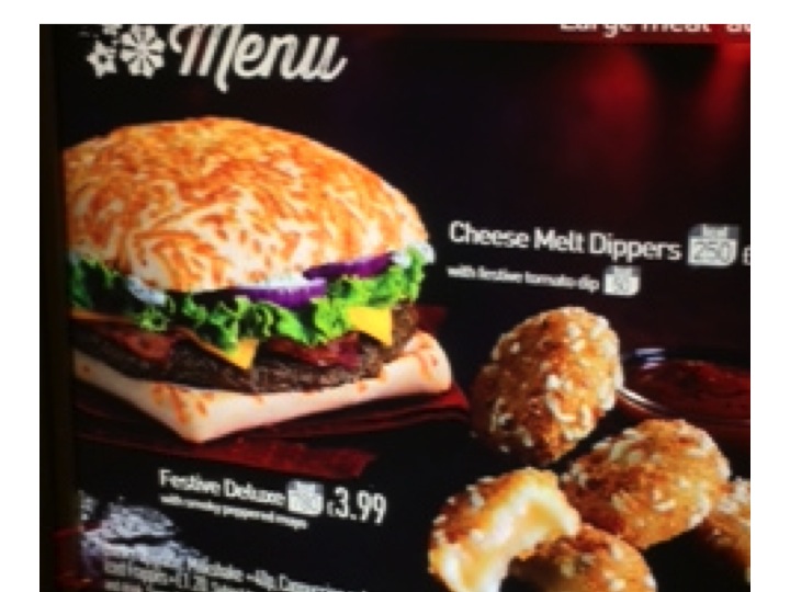
Diabetic ketoacidosis (DKA) is one of the more dramatic problems we deal with regularly on our acute medical unit.  Ryan didn’t even know he had diabetes before he came in, although the possibility had gone through his mind. He is nineteen and is studying economics at university. Last weekend he came home from college.  His mother immediately noticed that he had lost weight. She thought it was because he was not eating properly – but Ryan was insistent that he ate loads. He freely admitted his diet was mainly “rubbish” – lots of burgers and chips, and he drank loads of fizzy drinks (he does not like water).

Ryan would remove the green bits before eating this

Then on Saturday night he began to get abdominal pain and vomiting. Mum thought at first that the problem was that he had eaten some undercooked sausages and had too much to drink at a party at his friend’s house the day before.

On Sunday she and Ryan’s father became really worried. He was clearly more unwell, and his breath had a really odd smell – sweet and fruity.  And he was staggering to the bathroom every couple of hours to pass urine or vomit again, or both.  It was when he was too ill to get to the bathroom and peed in his bed that dad called the emergency out-of-hours doctor.  The GP realised straight away what was going on. Ryan was breathing deeply and heavily, dried saliva and vomit was crusted round his mouth and he was very dehydrated and sleepy.  She tested his blood for glucose – the reading was “hi” – off the scale. He was soon with us in hospital.

We have a protocol for dealing with DKA which involves lots of intravenous fluids – starting with normal saline, a continuous intravenous insulin infusion, and frequent and regular checks of blood acidity, glucose and potassium. We also look for any infection which might have precipitated the DKA.

There are lots of interesting and difficult questions to ask about type 1 diabetes, such as

–      what causes the pancreatic islet cells which make insulin to be destroyed?

–      why does this type of diabetes run in families?

But today I am going to concentrate on two easier questions:

–       why do type 1 diabetics lose weight before their disease is treated with insulin?

–       what causes ketoacidosis?

When I ask “why do diabetics lose weight?” the answer I often get is that they use up fat because their bodies cannot use glucose.

Glucose used to be thought of as a wonder food

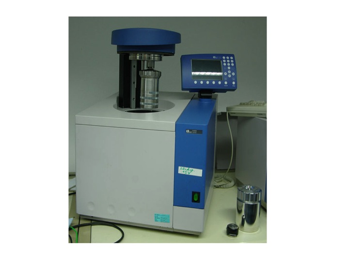
This answer does not really satisfy. When we eat food our digestive system is very good at extracting energy. We are all familiar with the calorie content of food. One calorie (really kilocalorie or kcal) is the energy required to heat a litre of water by one degree centigrade. The energy content of food, measured in kilocalories or kcal, can be accurately measured by putting the burger and chips in a device known as a bomb calorimeter.  This combusts the food with oxygen and measures the rise in temperature of the surrounding water bath. Let’s say the burger and chips, the three litres of cola, the cornflakes, milk and pork pie and other sundries that make up Ryan’s average daily food intake contains 2500kcal, if we were to put it in a bomb calorimeter.

bomb calorimeter - the bomb is the small silver cylinder- picture from wikimedia commons - by harbor1
bomb calorimeter – the bomb is the small silver cylinder- picture from wikimedia commons – by harbor1

Typically we absorb more than 95% of the available calories from an average meal. The only energy we cannot absorb is that in cellulose fibre*. Ryan does not eat much of that. Pretty much all of the 2500kcal he eats every day will be available as energy. Ryan is a fairly active young man. He uses 1500kcal a day just sitting studying or watching “man v food” on TV.  Walking, running, cycling and energetic evenings with his new girlfriend consume the other 1000kcal. So why is he now losing weight? Weight loss means loss of fat. This is stored all around our bodies as adipose tissue. Adipose tissue contains cells that essentially only contain a large blob of triglyceride (we’ll talk about that later). Triglyceride contains nine kcal of energy per gramme, so if fat disappears, the energy must be going somewhere. The second law of thermodynamics says that energy cannot be created or destroyed.

The best way of thinking about it is that energy is going in – 2500kcal/day – and energy is going out – basal metabolic rate and energy used for exercise – also 2500kcal/day. If these are balanced, then fat stores will not change. Clearly, if Ryan is losing weight and using up fat stores there must be an imbalance with this input and output equation. The problem is not with the input, he is eating loads and absorbing the energy. It’s not with his basal metabolic rate, that is normal. It’s not with energy expenditure with exercise – that has not changed. The energy loss is in Ryan’s urine – it is full of glucose.  Glucose has four kcal per gramme.  He is peeing out about 800kcal per day of energy – that is why he has lost weight. It is also why he is passing so much urine and become dehydrated. Glycosuria causes an osmotic diuresis – causing him to lose extra water and be very thirsty and drink lots of fluid. Unfortunately although the fizzy drinks have lots of sugar, it is going in one end and out the other. Once the vomiting started, he was not able to keep up with the input and became even more dehydrated.

Now the ketoacidosis. This has got very little to do with glucose – it has everything to do with fat metabolism. Insulin is the hormone which controls energy metabolism. When we eat, carbohydrate is turned into glucose. Blood glucose is monitored by pancreatic islet cells. pic If glucose is high, the islet cells release insulin.  Insulin has lots of important effects on how energy is used and stored.

When we are starved, with no carbohydrate intake, insulin levels drop and the lack of insulin:

1) promotes the conversion of glycogen (a glucose polymer stored in the liver) to glucose

2) keeps blood glucose levels acceptable by promoting the conversion of protein to glucose and

3) promotes breakdown of fat to provide energy.

The first two actions are important in keeping blood glucose high enough to maintain brain function– some brain cells absolutely rely on there being some glucose.

The third effect of low insulin –lipolysis – is also helpful in non-diabetics, breaking down fat to supply energy. Most tissues apart from the brain are very happy to get their energy from fat metabolism.

The problem comes when insulin is not just low, but completely absent. This never happens in non-diabetics. There is always enough insulin to keep fat breakdown under control. When insulin levels do go down to zero, in type 1 diabetics, fat starts to break down very quickly.

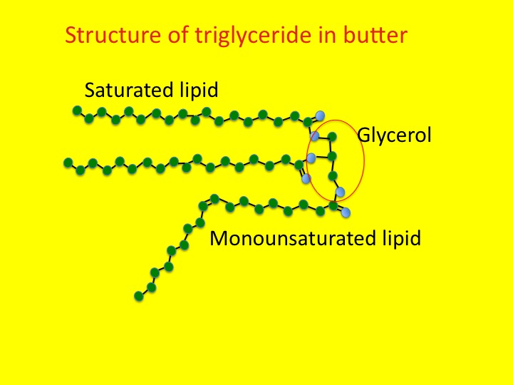
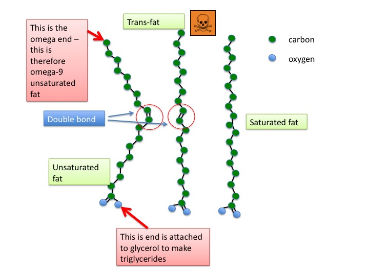
This process starts as lipolysis – the removal of the long fatty acid side chains from triglyceride. Triglyceride is an ester of fatty acid and glycerol. The glycerol backbone can be used to make more glucose (not that it is at all needed). Then the long-chain fatty acids are broken down, two carbons at a time. This is known as beta-oxidation.

triglyceride is first broken down by lipolysis, which removes the fatty acid chain - the fatty acid is then chopped up two carbons at a time by beta oxidation, attached to coenzyme A is then processed into carbon dioxide, water and energy by mitochondria
triglyceride is first broken down by lipolysis, which removes the fatty acid chain – the fatty acid is then chopped up two carbons at a time by beta oxidation, attached to coenzyme A is then processed into carbon dioxide, water and energy by mitochondria

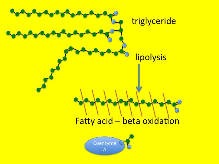
You will remember from previous posts (chest pain and horsemeat lasagne) that the carbon atom at the other end from the carboxylic acid group in a fatty acid is known as the omega carbon – hence omega 3,6 and 9 fatty acids which have double bonds in those positions. The carbon at the acid end of the molecule is the alpha carbon, and the next one along is, of course, a beta carbon. Hence beta-oxidation oxidises this carbon and removes an acetyl group from the fatty acid and attaches it to a Coenzyme A molecule. This can then be fed into the Kreb’s cycle and electron transport chain to convert it into carbon dioxide, water and nine kcal/gramme of energy in the form of ATP.

When insulin levels are very low, as was the case with Ryan, the rate of lipolysis and beta-oxidation are so fast that the Kreb’s cycle cannot keep up.

lipolysis is controlled by insulin levels in the blood - in non-diabetics insulin secretion does not drop below about 0.5 units/hr - type 1 diabetics can have no insulin and lipolyisis is unrestrained
lipolysis is controlled by insulin levels in the blood – in non-diabetics insulin secretion does not drop below about 0.5 units/hr – type 1 diabetics can have no insulin and lipolyisis is unrestrained

Fuel is being delivered at a faster rate than it can be burned. The irony of the situation is that there is plenty of fuel around in the form of glucose, but because it is not triggering an increase of insulin, the fat cells do not know this and keep on breaking down triglyceride. The only thing to do with all this broken down lipid is to make ketoacids (also known as ketones). In small amounts these are a quite useful alternative to glucose for energy production when we are starving – there are measurable amounts in blood and urine in healthy people if we do not eat for 12 hours or more, but low-level insulin secretion keeps the amount in check.Slide1

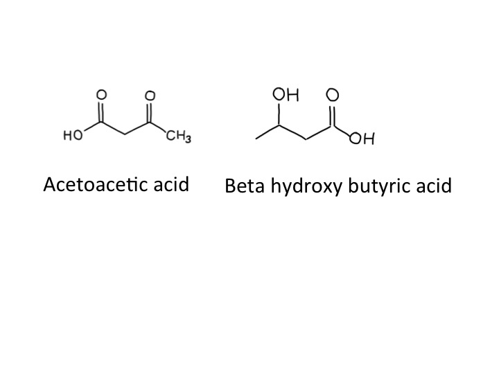
When ketoacids are made in huge amounts they cause problems because they are acidic. The two ketoacids we make are acetoacetic acid and beta-hydroxybutyric acid. Acetoacetic acid slowly, spontaneously degrades to make acetone and carbon dioxide,

acetone is commonly used as a solvent

which was why Ryan’s breath smelt sweet and fruity – like nail polish remover.  I think that beta-hydroxy butyrate is not technically a ketone – but it is an acid. I don’t think it is necessary to make a fuss about this (but the pedant in me forced me to mention this).  Acidosis makes people ill – our bodies are designed to work with a blood pH of between 7.35 and 7.45. Ryans blood pH was 7.04 when we first tested it. Many of the enzymes in our body just don’t work properly when the blood is too acid – when it becomes too severe acidosis can cause drowsiness and then coma and then death. The other problem is that the physical stress caused by ketoacidosis and dehydration from high glucose result in the release of glucocorticoids (cortisol) and adrenaline from our adrenal glands, both of which further encourage lipolysis and the formation of more ketoacids. A potentially lethal viscious cycle.

How does neutral fat make acidic substances? Well, acidity is not like water, sodium or energy where what goes in must come out. We can drink lots of acidic vinegar and it will not cause any change in blood acidity. The easiest way to look at it is that when we make small, charged molecules from large uncharged fat molecules, the hydrogen atoms are happy to give an electron to the anion (such as acetate) and drift off as a positive hydrogen ion – ie increase acidity.

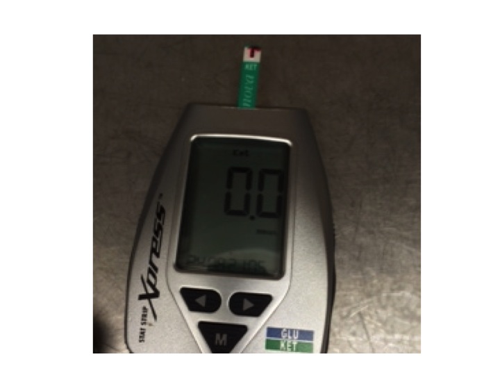
We always used to rely on measuring urine ketones, but now have a blood ketone meter which is much better at quantifying the amounts in the blood of patients with ketoacidosis. Ryan had a level of 9.6mM when he came in. By the next day he was sitting up, eating lunch, with a level of 0.3mM.

blood ketone meter - I measured my blood just after breakfast - that was why the reading was zero
blood ketone meter – I measured my blood just after breakfast – that was why the reading was zero

We used to use a sliding scale to treat patients with ketoacidosis. This meant that the higher the blood glucose, the more insulin we gave. It is quite clear from what I have said about what causes ketoacidosis that this was not a sensible strategy. We now give enough insulin (>6units/hr) to completely supress lipolysis until the level of ketones in the blood becomes normal, and usually have to give extra glucose to prevent blood sugar levels going too low.

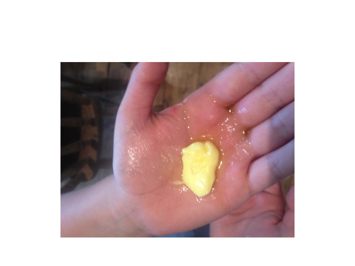
Now to the food link – butter.  As previously discussed, butter is mainly made from triglyceride. It is yellow because it contains vitamin A, or carotene. This helps prevent it becoming oxidised, or rancid. Slide4 
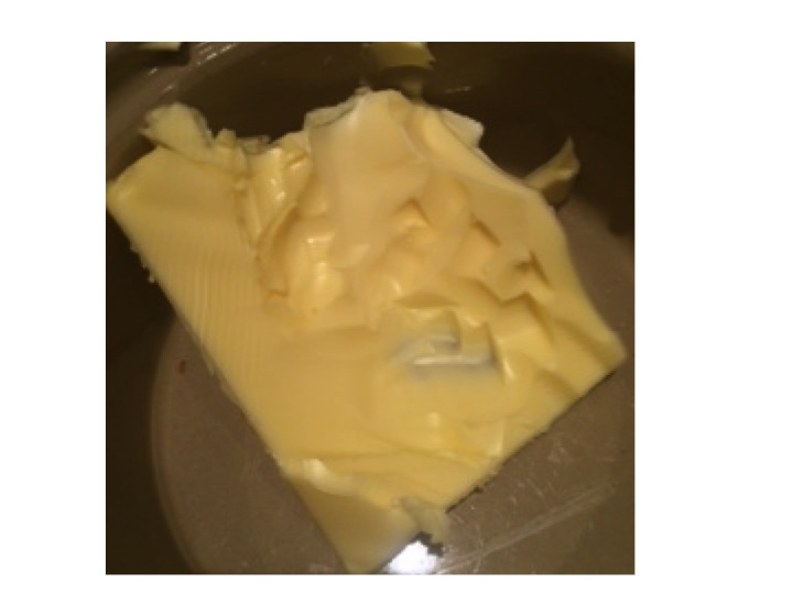
When butter does goes rancid, it is because the fatty acid chains become oxidised by bacteria, in a process similar to beta-oxidation. One of the main oxidation products of butter is called butyric acid. And, amazingly, the reason butyric acid is called that is because it was first identified in rancid butter – Latin for butter is butyrum (cow-cheese). The simple hydrocarbon with four carbon atoms then became known as butane, again named from butter/butyrum. Not sure why methane, ethane or propane are called that. If I find out I will let you know in a future post.

*cellulose is a glucose polymer made by plants to provide structural support – when it is eaten as food it is known as fibre – it burns well in a calorimeter but cannot be used by humans to provide useful calories – but can be used as an energy source by cows, which, of course, provide us with butter.  Ryan likes butter but he does not like fibre. The diabetes dieticians are trying to change that.  I wonder if he will be eating brown-bread toast now? (see my very first post – toast and diabetes).


gallstones and mayonnaise

A few weeks ago we admitted Sylvia. She had been feeling unwell for a some days, with fever and episodes of uncontrollable shaking. What really alarmed her was when she turned yellow, and then developed abdominal pain and vomited a couple of times. Sylvia is forty eight, and had always been healthy. She rowed with the local club twice a week and kept her weight under control. For the past year or so she noticed that she got pains in her abdomen after eating fatty foods such as fish and chips, and now avoided foods like that. It was her son who noticed she was yellow – he then phoned Sylvia’s GP. The duty doctor came and confirmed that she was indeed jaundiced, feverish and unwell, and persuaded her that she needed to go to hospital.

these people look jaundiced - in fact they are not - the whites of their eyes are white - this image is copyrighted but Wiki thinks its ok to use it so I'm hoping its ok for me to do so
these people look jaundiced – in fact they are not – the whites of their eyes are white – this image is copyrighted but Wiki thinks its ok to use it so I’m hoping its ok for me to do so

We did all the usual things for someone with suspected biliary sepsis. Took blood cultures and routine blood tests. These tests confirmed she had elevated bilirubin and transaminases, increased alkaline phosphatase, as well as a high neutrophil count and CRP.  We gave her intravenous fluids, anti-emetics and antibiotics. We were able to get an ultrasound within a couple of hours. Ultrasound is really good at looking at the liver and biliary system in jaundiced patients – better than CT.

The ultrasound showed that Sylvia had a gallbladder full of gallstones and her common bile duct was dilated. It seemed likely that she had a stone blocking the flow of bile from the liver to the duodenum, and that the stagnant bile had become infected with bacteria causing ascending cholangitis and liver inflammation.

bile is made in the liver - it is stored in the gallbladder and squirted into the small intestine after a fatty meal, along with pancreatic secretions - Sylvia's gallstone was impacted in the sphincter of Oddi
bile is made in the liver – it is stored in the gallbladder and squirted into the small intestine after a fatty meal, along with pancreatic secretions – Sylvia’s gallstone was impacted in the sphincter of Oddi

The questions I want to answer this week are:

Why did Sylvia turn yellow?

What are gallstones made of and why do they form?

Why is there an increase in alkaline phosphatase in the bloodstream when the biliary system is obstructed?

The last of those questions is the hardest, but I will make some conjectures, and  hope that others who know more about it will correct me.

Jaundice is fairly straightforward. It is caused by the accumulation of bilirubin, which is a greenish-yellow colour. Bilirubin is made from haem, the central working part of myoglobin, but most bilirubin comes from the breakdown of haem in the haemoglobin from red blood cells.

So why is haemoglobin red? It is not only – or even mainly – the iron that makes haem red, but the porphorin ring that surrounds the iron. This large ring is made of four smaller pyrrole rings.

Pyrroles are often coloured – the dye indigo has two pyrrole rings and red acrylic paint often contains the dye pyrrole-red. Melanin, the pigment that gives skin and hair its colour is brown or black or red because it has a related structure – indole – a pyrrole stuck to a benzene ring. Without this pyrrole ring we would be like albinos, but without pink skin and eyes.  As we shall see, we would also produce beige poo and colourless urine.

this is a pyrrole - all pyrroles are highly coloured, and are in most of the things which make people the colour they are
this is a pyrrole – all pyrroles are highly coloured, and are in most of the things which make people the colour they are

Red blood cells last on average 120 days before they wear out and have to be recycled.  As we have five litres of blood and in each litre there is at least 120 grammes of haemoglobin, it means we have to make, and break down, five grammes a day.  About a teaspoonful.  This all happens inside macrophages in the spleen and liver, as they try to salvage useful parts of the red cells to make new things.

Macrophages are similar to neutrophils but slightly smarter.  Some are good at host defence: they come to the scene after the neutrophils have done their job, do the forensics, and find out what the germs are made of. They then tell the lymphocytes how to make antibodies to protect us in the future.

Other macrophages are good at clearing up the mess, breaking down dead tissue and making things neat and clean.  The spleen and liver macrophages that deal with old red cells are the latter kind. When they take red cells apart, they separate haem from globin, and then take the iron out of the haem.  Next, the porphorin ring is broken to form bilirubin – its molecule of linked pyrrole rings looks like four beach huts in a row.

haem is broken down by the enzyme haem oxygenase to form bilirubin

The bilirubin is then thrown away (I don’t know why it can’t be re-used).  It attaches to albumin and becomes water soluble after conjugating in the liver with a sugary molecule called glucuronate.  The conjugated bilirubin is then transported into the biliary system, stored in the gallbladder, and squirted into the duodenum when we eat fatty food.  In the bowel, it is converted to urobilinogen, which makes poo brown.  Some is reabsorbed and excreted in urine, which makes urine yellow.Slide2

Sylvia had noticed that her poo had changed colour – it had become putty-coloured – and her urine had become much darker. That is because conjugated bilirubin was not getting into her intestines and instead some had leaked into her bloodstream and was appearing in her urine. The whites of her eyes and skin had become yellow because of the very high level of conjugated bilirubin in her blood.

Now to the gallstones.  In Western countries they are most commonly made of cholesterol.  I have already talked about the importance of cholesterol in keeping cell membranes rigid and non-leaky.  Cholesterol is made in large amounts by the liver and transported to other cells gift-wrapped as LDL cholesterol.  It is also used to make bile salts.  It only needs a minor modification of the basic chemical structure of cholesterol to make bile salts – the main component in bile which makes it work – the main function of bile salts is to help us digest dietary fat.

structure of cholesterol

Bile salts are detergents.  Just like washing-up liquid, they emulsify fats, breaking large fat globules into smaller micelles which do not stick to each other, or anything else. Cholesterol is essentially very insoluble in water, or hydrophobic.  When an organic acid group is added – to make bile salts – it gains a hydrophilic or water-attracting group. This is just like soap – a long lipid chain with an organic acid group at the end.  The hydrophobic lipid part is embedded in the tiny fat globule and the hydrophilic groups stick outside in the watery medium of the intestinal contents.

bile acids are a minor modification of cholesterol - hydrophilic OH  and COOH  groups are added to make it into a detergent
bile acids are a minor modification of cholesterol – hydrophilic OH and COOH groups are added to make it into a detergent

As well as bile salts, the liver secretes phospholipids into bile – principally phosphatidylcholine. This, with cholesterol, is the main stuff cell membranes are made of.  It is also an emulsifying agent, and is used extensively in the food industry to keep fats in suspension.

So, when we eat fat, the small intestine detects it and releases the hormone cholecystokinin (CCK).  This causes the gallbladder to contract to send bile into the duodenum, and also makes the pancreas release enzymes, including lipase.  CCK also has some very interesting effects on our brains, making us feel less hungry, and curiously, opposing the effects of opiate drugs.  The bile and pancreatic secretions are both delivered into the duodenum through the same tube – see diagram. The detergent bile salts, emulsifying phospholipid and pancreatic lipase are designed to work together to digest fat.

the hydrophilic parts of cholesterol are all on one side of the molecule - this allow it to act as a detergent - it works well to disperse fat into small globules so that pancreatic lipase can work on it and break it down to fatty acids and monoglyceride
the hydrophilic parts of cholesterol are all on one side of the molecule – this allow it to act as a detergent – it works well to disperse fat into small globules so that pancreatic lipase can work on it and break it down to fatty acids and monoglyceride

The bile breaks it up into tiny globules and the lipase breaks triglyceride into fatty acids and monoglyceride. These are transported across the intestinal mucosal cell membrane and then the triglyceride is put back together again. Seems a daft system, but it clearly works. The triglyceride is taken away from the intestine not in the portal blood, like most other substances absorbed by the gut, but in the lymphatic system in the form of chylomicrons – small fatty globules covered by a layer of phospholipid. The chylomicrons travel up the lymphatic vessels to emerge into the circulation just below our left clavicle. This means that it avoids passing straight away through the liver. If blood is taken soon after a fatty meal and centrifuged, it will have a milky appearance because of the large amount of chylomicrons.

So why do gallstones form? As well as bile salts, unmodified cholesterol is also secreted into bile. It is only kept soluble by the bile salts and phospholipid – the ratio of cholesterol to bile salts and phospholipid is therefore important. When it goes wrong, cholesterol precipitates out and forms solid stones like those that caused all Sylvia’s recent problems – a bit like when I try to make mayonnaise and it curdles – the fat becomes un-emulsified and separates out.

In some parts of the world, gallstones are made from bile pigment – bilirubin. This can be due to increased red blood cell breakdown resulting from abnormalities such as sickle cell disease, but it’s also common in East Asia, for reasons which are not clear.  A good, comprehensive analysis of why gallstones of all types form can be found at:

http://gastro.ucsd.edu/fellowship/materials/Documents/Gallstones/pathogenesis%20gallstones.pdf

Now the alkaline phosphatase. Bile is alkaline.  Alkaline phosphatase is an enzyme that removes phosphate groups in alkaline conditions.  When bile flow is blocked – by stones, as in Sylvia’s case, or for any other reason – the liver makes much more alkaline phosphatase, some of which appears in the blood.  Bones use a similar alkaline phosphatase to rearrange phosphate groups to make hydroxyapatite – the hard stuff bone is made from.  Most of the alkaline phosphatase normally in our blood is the bone sort.

There is plenty of information about whether alkaline phosphatase is from bone or liver, but very little I can find that suggests why the liver should make this enzyme when the biliary system is blocked.  I think a clue here is that neutrophils also have alkaline phosphatase in their granules, which help kill bacteria.  This is in addition to the myeloperoxidase and esterase mentioned in earlier posts.  Why should a phosphatase be damaging to bacteria?  The answer may well involve techoic acid, an important reinforcing molecule in some gram-positive bacteria.

cartoon of gram positive bacterial cell wall - techoic acid is thought to inhibit the action of lysozyme and protect the peptidoglycan from being broken down - techoic acid contains phosphate groups which may be removed by alkaline phosphatase
cartoon of gram positive bacterial cell wall – techoic acid is thought to inhibit the action of lysozyme and protect the peptidoglycan from being broken down – techoic acid contains phosphate groups which may be removed by alkaline phosphatase

In these microbes, techoic acid strengthens the peptidoglycan cell wall and inhibits the action of lysozyme, yet another enzyme made by neutrophils.  Lysozyme is designed to break some of the sugar-sugar bonds in peptidoglycan – so getting rid of the techoic acid would surely be helpful.  We know that alkaline phosphatase is effective in breaking up techoic acid – see, for instance:

http://jb.asm.org/content/102/3/747.short

Maybe the liver is producing this anti-bacterial enzyme to help prevent infection when the flow of bile slows down.  Unfortunately, this won’t work with gram negative organisms such as E.coli because they don’t make techoic acid – and it was a gram negative bacterium we grew from Sylvia’s blood culture – the germ that was causing her ascending cholangitis.

Soon after she came in, Sylvia had an ERCP – an endoscopic retrograde cholangio-pancreatoscopy.  The endoscopist managed to remove the impacted gallstone from her ampulla of Vater, and she quickly recovered.  A week later she had her gallbladder removed, including the stones, and now is quite well – she can even eat fish and chips.

Now to the food link: mayonnaise.

French mayonnaise - made with olive oil and mustard
French mayonnaise – made with olive oil and mustard

This is an emulsion of lipid, such as olive oil, in an aqueous (watery) medium – vinegar or lemon juice.  The emulsifying agent is raw egg yolk, which – just like bile – contains lots of phospholipid – phophatidylcholine and is also rich in cholesterol.  In France, mustard is also added.  Mustard seed contains mucilage, a gooey stuff that some plants produce made of sugar polymers. (Thanks again to Harold McGee). These also act as emulsifiers and further thicken the mayonnaise, and give it a better taste. When I make cheese sauce, I use a sugar polymer (flour) to achieve a stable emulsion between butter and milk – no doubt the phospholipid in the milk helps too.  It seems likely that in the small intestine, the mucus from the stomach, a similar gloopy sugar polymer, probably has a similar effect to mustard seed mucilage to help with fat emulsification.

Chest pain and horsemeat lasagna

A heart attack, or myocardial infarction to give it the proper name, is still a very common cause of death in the UK. Those unfortunate patients with severe heart attacks don’t even touch the ground, going straight to the cardiology department to have the offending blood clot removed from their coronary artery. In the medical admissions unit we also see a lot of patients with chest pain who have less severe heart attacks.

Today we admitted Toby, age 57, who had bad episode of chest pain which woke him up at 4am. The pain was so bad it made him vomit. It was a like a heavy pressure on the front of his chest. His wife woke up and noticed that he looked really pale and ghastly and realised there was something seriously wrong,

“No, its just a bit of indigestion dear, you go back to sleep, I’ll be OK”,

he said, at the same time also knowing that this was not the case. Of course she did the sensible thing and called the ambulance. When the ambulance men arrived they gave him morphine, oxygen, nitroglycerine and some aspirin tablets to chew. They did an EGC and sent the result to our ED department – not normal but not a major heart attack.

When he arrived we measured the level of troponin (a substance that indicates heart muscle damage) in his blood – it was increased to over 200 – the normal upper limit in our hospital is less than 14. He had an NSTEMI, that is a non-ST elevation myocardial infarction. That meant the damage was probably caused by a partial rather than complete blockage of his coronary artery.

What causes the pain with heart attacks? When a coronary artery is blocked, or partially blocked, the bit of heart muscle it normally supplies gets starved of oxygen. It needs oxygen to make energy in the form of ATP to keep on beating. In the hope that oxygen will be soon restored, the cardiac cells take out a pay-day loan.

The cells that have spent all their oxygen convert glucose into lactic acid to produce four ATP molecules. Normally, when oxygen is available a glucose molecule produces about 30 molecules of ATP. Lactic acid produced in the abscence of oxygen lowers the pH (more acid) and stimulates sensory nerve cells – this is perceived as pain.

Bad pain. Visceral pain. The sort that makes you vomit and makes think you are going to die. Payday loans are always costly. About half of the patients with full-thickness STEMIs who die, do so before they get to hospital. This is mainly because the lack of oxygen and metabolic upset caused by the blocked coronary artery causes ventricular fibrillation – a fast and totally chaotic electrical activity which results in no useful heart function and death in a few minutes if not terminated by an electric shock.

So why did Toby get a blocked coronary artery? Sorry, I now realise this is going to be a longer blog than usual.

I think it is useful to know that traditionally, the Japanese suffer from far less coronary artery disease than those in the US or northern Europe. By far less I mean about 30%. This is a huge difference. If we were like the Japanese we could dramatically reduce the number of patients admitted with coronary artery disease and probably reduce our cardiac department costs by over a half.

There is plenty of evidence that it is what the Japanese eat which protects them from coronary heart disease. The traditional Japanese diet has a lot of fish and vegetables. When they move to the US and eat hamburgers, fries, cookies, and donuts their rate of coronary heart disease soon matches the rest of the US population.

Slide1

What is also interesting is that those in Southern Europe eating a “Mediterranean diet” tend to have much lower rates of heart disease than in Northern Europe (although not as low as Japan). Their diet also has quite a bit of fish, but also has a lot of olive oil, pasta, and bread.

I think what we do know is that saturated fats are quite bad, trans- fats are very bad, and fish is good. What follows is an attempt to explain why, with a mix of accepted orthodoxy and some speculation. If you think my speculation is way out of line I hope you will tell me.

The traditional Northern European diet relies heavily on wheat, with supplementation by milk and dairy products, particularly in the winter. Northern Europeans have a mutation which means that they can drink large amounts of milk as adults. Infants, of course, can drink milk, but to be able to absorb the milk sugar lactose they need the enzyme lactase to break the disaccharide into glucose and galactose. This enzyme is conveniently situated in babies on the surface of intestinal cells. For most of the world’s population, the lactase enzyme disappears at age 2 or 3, meaning that if they drink more than a cup full of fresh milk they will get diarrhoea. The lactose is not absorbed but goes to feed bacteria in  the colon who have a party. Normally these bacteria see only the leftovers from our diet such as cellulose fibre. The result of the celebrations is a lot of gas, abdominal cramps and diarrhoea, otherwise known as lactose intolerance (not to be confused with milk allergy). The ability to drink milk without these problems as an adult is known as lactase persistence. This mutation has allowed humans to colonise Northern Europe and survive in the Winter by drinking milk, and more recently eating dairy products. This is a link to a fascinating blog post all about lactase persistence and a map showing which adults in the world can drink milk, and those who can’t.

http://sci.waikato.ac.nz/bioblog/2010/06/the-genetics-of-lactase-persis.shtml

The wheat we traditionally grow in Northern Europe is not good for making bread. It does not have enough of the springy gluten protein to allow large bubbles to form in the rising dough. The bread turns out heavy and unappetising. Having said that, it was the staple food of most  people in the UK until relatively recently. What English flour is really good for is making pastry and biscuits (cookies). For that you need low gluten (weak) flour and hard fat, such as butter or lard – or indeed, suet. Butter has always been plentiful in England because of the huge dairy industry – turning milk into butter preserves most of the calories and stops it spoiling so quickly.

Pastry cooks know that it is important to keep your hands cold when making pastry – the fat must stay hard when it is mixed with the flour to give the pastry that wonderful crumbly, delicate, tasty and satisfying feel in your mouth. Try to make pastry with olive oil or sunflower oil and you will get a tough, chewy and very disappointing result. I presume it is because the liquid oil seeps into the starch grains of flour, making them stick together, rather than coating them and keeping them apart during cooking (any food scientists out there?)

cakes

cakes made by my family today – from butter, not margarine

The problem is that butter contains mostly saturated fat. You can generally tell how saturated a fat is by how hard it is at room temperature. Butter is slightly more saturated than lard, but less saturated than suet, the fat surrounding kidneys. Olive oil is monounsaturated – it is normally liquid but will solidify in the refrigerator. Sunflower oil is polyunsaturated – it will stay liquid even when quite cold.

Saturation depends on how many double bonds there are in the long fat chains which make up triglyceride. Triglyceride is the way fat is stored in nature – in plants, insects, fish and mammals, including humans. Here is picture of a typical triglyceride in butter:

Slide1
green dots are carbon atoms, blue are oxygen

You will see that it is made of three long chains of carbon molecules, joined onto a backbone of glycerol – a 3 carbon molecule. Why is all fat in nature stored with this arrangement? Why not a backbone of 2 carbons, or 4 or 20? I don’t know.  Answers please.

The reason why saturated fat is solid at room temperatures is because the long fat chains are straight.  Unsaturated fats have a double bond C=C somewhere in them which makes the chain bent.

Slide6

Bent chains don’t line up next to each other so well, and are more reluctant therefore to solidify and stay liquid at lower temperatures than saturated fats with the same length of carbon chains.

Saturated fats come mainly from mammals. Unsaturated fats come from plants and fish. There is a reason for this. Plants have to endure cold temperatures. Fish live in cold water and are poikilothermic – their bodies are a similar temperature to the water. Mammals stay warm, with a core temperature of 37 degrees. If you take a lump of butter and hold it in the palm of your hand it will melt.

Slide2

So, in the cow the fat is liquid. If you buy a fish and put it in the fridge, it will stay bendy, whereas a pork chop will go stiff. We all need to have liquid fat so we can easily move it around. Have a feel of that fat under your abdominal skin – it is liquid. That’s because it is 37 degrees Celcius. If it was at room temperature it would be at least semi-solid.

One of the reasons that butter, or cow fat, is so saturated is because of the strange digestion system in cows. All the grass they eat goes first into an enormous biodigestor called the rumen. The rumen is huge. Over 200 litres – bigger than a person. Here cellulose is broken down by bacteria to simpler sugars. This fermented grass is then regurgitated back into the cow’s mouth, chewed some more, then sent down a different tube to other stomachs, ending up in the abomasum, which is similar to the human stomach when all the goodies in the grass are in a form which can be absorbed. The problem is that any unsaturated fat in the grass will be chemically reduced in the rumen to saturated fat. This fat goes to make milk and butter.

The rumen of a cow is huge - it fills up much of the inside of the animal
The rumen of a cow is huge – it fills up much of the inside of the animal. Unsaturated vegetable fats are reduced to saturated fats in the rumen.

I must mention trans-fat here. Butter is relatively plentiful in Northern Europe, but has always been a quite expensive luxury. Napoleon offered a prize to anyone who could invent a cheaper substitute for butter. Margarine was originally made from beef fat, but it later developed as a way to make cheaper oils solid by reducing the C=C double bonds into simple C-C bonds by reaction with hydrogen and nickel catalyst. Hydrogenation of unsaturated vegetable fat to make it saturated will make it hard and good for making cakes and pastry. Unfortunately it  can also cause the formation of C=C bonds in the trans- formation rather than the normal cis- formation. Trans- double bonds do not cause the lipid chain to be so bent. The straight chains are the reason that margarine is solid at room temperature.

Napoleon never invaded England, he instead invented a fiendish foodstuff which over the years has probably killed a very large number of them. Trans fats are now banned in the US and most European countries. Margarine manufacturers have now devised other ways to make liquid fat solid without the formation of trans- fats.

So why is saturated fat, and even more, trans- fat bad for us? Why does it cause coronary arteries to block up? The reason is that it increases the amount of cholesterol in our bodies, in particular the low density lipoprotein cholesterol – LDL.  You might think that the reason why saturated and trans-fats increase LDL cholesterol is well known. Well, I read the book below and could not find the answer. Not online either. The following is my best guess.

This book tells you almost all you need to know about fats.

We have to take another aside here to talk about cholesterol. This is completely different from triglycerides. Cholesterol is an amazing molecule. It is only made by animals – vegans get no cholesterol in their diet, but luckily they can make it. I say luckily, because it is a totally vital substance which has many very important uses in our bodies. The one we are going to talk about here is its function as a constituent of cell membranes. All the cells in our bodies are surrounded by a membrane made of phospholipids – these molecules are similar to triglycerides but have just two fat chains attached to a phosphate group.

structure of cholesterol
structure of cholesterol – not at all like triglycerides

Membranes made of just phospholipids do not do all the things we need a membrane to do. It will not be strong enough, rigid enough, and will be too leaky. Small molecules like sodium ions, water and glucose would be able to leak through and cells could not work at all.

Cholesterol is incorporated into the phospholipid membrane to make it stiffer and less leaky

So cholesterol is used in large amounts to make the membrane stiffer and less leaky. In many membranes there is as much cholesterol as phospholipid. All cells can make cholesterol, but it is a real pain. There are over 30 enzymatic steps needed to make cholesterol from glucose. An analogy might be clothes. We could all make our own clothes if we needed to – buy the cloth, cut it up and sew all the pieces together. But we’d rather buy them from a shop – much easier. Similarly for the average cell – it needs cholesterol to incorporate into its membranes – it could make it,  but there is a ready supply in the bloodstream in the form of LDL cholesterol particles made by the liver – you don’t even have to order it – it is on tap all the time. The liver is good at a whole range of things, making cholesterol is one of them. It sends the cholesterol in little gift-wrapped packages, covered with a special protein coat, continually into the bloodstream to be picked up by cells around the body. Cells which need cholesterol make a special “docking port” on their surface to trap the LDL particle, take it inside the cell and use the cholesterol to beef up and stiffen up its membranes. These docking ports are called LDL receptors. Seems like a good system.

Gift-wrapped LDL particles are taken up by cells to provide cholesterol for cell membranes

The problem is that when we eat too much saturated, hard fat, or trans- fat, these straight lipid chains get incorporated into the cell membranes. Because the chains are straight, the membranes are more rigid and less leaky. The cells therefore don’t need so much cholesterol to make the cell membranes nice and stiff and work properly. So the liver is pumping out LDL cholesterol, and the cells don’t want it.

It seems that the liver does not have a good system of knowing how much LDL is needed. It keeps making LDL cholesterol and it hangs around, like an unemployed teenager. Then it inevitably gets mixed up with the wrong company – oxidising agents. Oxidation (from enzymes such as myeloperoxidase – see previous post on phlegm and horseradish) changes the LDL particle in a bad way, and it inevitably gets picked up by the police (see previous blog on asthma). Here the police are macrophages. They, for some unknown reason take the delinquent LDL particles and imprison them in blood vessels, just below the lining cells or endothelium. With time (Toby has been eating pies and cookies with enthusiasm for more than 50 years) these bad cholesterol prisons become fill up with inmates. They make a lump on the inside of the artery. The lump slowly gets bigger and bigger, making the hole down the middle of the artery smaller and smaller. When Toby woke up at 4am with chest pain, the surface of the lump, or atheromatous plaque to give its proper name, ruptured. Blood flow to part of his heart was cut off by platelets trying to mend a hole which was not really there.

When a blood vessel is damaged, it is normally vital to plug up the hole. The first defence against bleeding to death from a hole in your blood vessel are platelets. These are tiny little bags made from bits of cells in the bone marrow called megakaryocyes. Platelets continually bud off from megakaryocytes and are packed with all the technology needed to find and mend holes in blood vessels. They have no nuclei, so are really not cells, more like pre-programmed drones which cruise round the bloodstream looking for holes.

“Your mission, should you choose to accept it*, is to go out and look for holes in blood vessels. You will know there is a hole because you will come into contact with collagen – that is sure-fire evidence of a hole. When you find the hole you will go into activate mode. You will change from smooth pebble shape to crazy octopus-with-lots-of-legs shape and you will stick onto the collagen with your sticky tentacles. You will immediately summon your platelet colleagues by making thromboxane and ADP. They will come and stick to you to help staunch the flow. If you detect thromboxane or ADP made by your fellow platelets you will go into activation mode at once and join your brave colleages until the hole is repaired. Good luck, and remember it is your mission to Save The Human Race”

When platelets encounter collagen they activate and change shape. They stick together with the sticky glycoprotein IIb/IIIa. Activated platelets release thromboxane and ADP to tell nearby unactivated platelets to come and join the party.

Thromboxane is made from the cell membrane phospholipids of platelets, from a very unsaturated fat called arachadonic acid. The enzyme which makes thromboxane from arachadonic acid is called cyclo-oxygenase. The reason why the ambulance crew asked Toby to chew two aspirins is because this drug very effectively inhibits cyclooxygenase enzyme and prevents platelet activation by preventing thromboxane production. He was given clopidogrel when he arrived in ED. This prevents platelets from responding to ADP released from their colleagues by blocking the ADP receptor.

So the problem was that the atheromatous plaque, or fatty lump in his coronary artery had ruptured. This activated the platelet drones which were zooming by. Collagen is found in the wall of blood vessels, but is covered by endothelial cells, the cells lining blood vessels. When the plaque ruptures, as well as a soup of cholesterol and macrophages, there is a lot of collagen. The platelets go crazy – a feeding frenzy fuelled by massive release of thromboxane and ADP. Before long the whole coronary artery is full of activated platelets. If this happens in a large coronary artery the result is a STEMI: full thickness cardiac damage. Toby was lucky, his clot only caused partial thickness damage, but was still very painful and will mean that his heart will not work quite as well as it did.

Why are trans-fats worse than ordinary saturated fats? I don’t think there is a definite cause known, but it may be simply that once the straight trans- fat is incorporated into cell membranes it is more difficult for the normal metabolic process to add further cis- double bonds and elongate the lipid chain into a bent and more liquid form. This means that cells need even less cholesterol to make their membranes stiffer, and LDL cholesterol is even more redundant and superfluous to requirements.

We have understood about unstable atheromatous plaques only relatively recently. A really important contribution to our understanding was made by Michael Davies who died in 2003. There is a short and very understandable youtube video showing him dissecting a coronary artery from a patient who died from a STEMI.

See:

http://www.youtube.com/watch?v=F0Y5hss1tZQ

The clot in the coronary artery in the video is made of white thrombus – basically activated platelets sticking together, with a mission to save lives but causing death. Drones can be problematic. Michael Davies discovered why about 15% of people in the US and UK die. He has not even got a Wikipaedia page and there are no photos of him on Google images– can someone sort this?

I mentioned that fish is good to prevent heart disease. The Japanese eat lots of it. I have started to eat more fish. Fish oil has an extra double bond near the end of the lipid chain – 3 bonds from the end – omega 3. (Omega is the letter at the end of the Greek alphabet, so if we are counting bonds from the end of the lipid chain the omega notation is used). Now if we eat fish, the lipid chains in our membranes have an extra cis- double bond near the end. When our platelets use these lipids to make thromboxane, a slightly defective form is produced with the extra double bond. This is known as TXA3  rather than the normal TXA2. TXA3 does not work as well as TXA2 and platelets cannot stick together so fast. This is a good thing if you have a ruptured atheromatous plaque in your coronary artery.

Now the food link – dodgy lasagne. Horses do not have a rumen. They eat and digest vegetable matter in a different way from ruminants like cows and sheep. They first take out the goodness they can in a normal stomach, then ferment the residue in a huge caecum, which breaks down the cellulose into simpler, digestible molecules. They are known as hindgut fermenters. This means that the vegetable lipids are absorbed in the stomach and stay unsaturated and horse meat contains fat which is much healthier than cow or sheep fat. Ironically (given the recent scandal about horsemeat in lasagne), horsemeat is therefore better for us than beef.

*They don’t really have a choice.