Tag Archives: pancreas

gallstones and mayonnaise

A few weeks ago we admitted Sylvia. She had been feeling unwell for a some days, with fever and episodes of uncontrollable shaking. What really alarmed her was when she turned yellow, and then developed abdominal pain and vomited a couple of times. Sylvia is forty eight, and had always been healthy. She rowed with the local club twice a week and kept her weight under control. For the past year or so she noticed that she got pains in her abdomen after eating fatty foods such as fish and chips, and now avoided foods like that. It was her son who noticed she was yellow – he then phoned Sylvia’s GP. The duty doctor came and confirmed that she was indeed jaundiced, feverish and unwell, and persuaded her that she needed to go to hospital.

these people look jaundiced - in fact they are not - the whites of their eyes are white - this image is copyrighted but Wiki thinks its ok to use it so I'm hoping its ok for me to do so
these people look jaundiced – in fact they are not – the whites of their eyes are white – this image is copyrighted but Wiki thinks its ok to use it so I’m hoping its ok for me to do so

We did all the usual things for someone with suspected biliary sepsis. Took blood cultures and routine blood tests. These tests confirmed she had elevated bilirubin and transaminases, increased alkaline phosphatase, as well as a high neutrophil count and CRP.  We gave her intravenous fluids, anti-emetics and antibiotics. We were able to get an ultrasound within a couple of hours. Ultrasound is really good at looking at the liver and biliary system in jaundiced patients – better than CT.

The ultrasound showed that Sylvia had a gallbladder full of gallstones and her common bile duct was dilated. It seemed likely that she had a stone blocking the flow of bile from the liver to the duodenum, and that the stagnant bile had become infected with bacteria causing ascending cholangitis and liver inflammation.

bile is made in the liver - it is stored in the gallbladder and squirted into the small intestine after a fatty meal, along with pancreatic secretions - Sylvia's gallstone was impacted in the sphincter of Oddi
bile is made in the liver – it is stored in the gallbladder and squirted into the small intestine after a fatty meal, along with pancreatic secretions – Sylvia’s gallstone was impacted in the sphincter of Oddi

The questions I want to answer this week are:

Why did Sylvia turn yellow?

What are gallstones made of and why do they form?

Why is there an increase in alkaline phosphatase in the bloodstream when the biliary system is obstructed?

The last of those questions is the hardest, but I will make some conjectures, and  hope that others who know more about it will correct me.

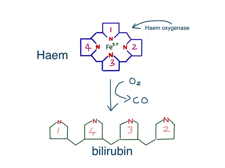
Jaundice is fairly straightforward. It is caused by the accumulation of bilirubin, which is a greenish-yellow colour. Bilirubin is made from haem, the central working part of myoglobin, but most bilirubin comes from the breakdown of haem in the haemoglobin from red blood cells.

So why is haemoglobin red? It is not only – or even mainly – the iron that makes haem red, but the porphorin ring that surrounds the iron. This large ring is made of four smaller pyrrole rings.

Pyrroles are often coloured – the dye indigo has two pyrrole rings and red acrylic paint often contains the dye pyrrole-red. Melanin, the pigment that gives skin and hair its colour is brown or black or red because it has a related structure – indole – a pyrrole stuck to a benzene ring. Without this pyrrole ring we would be like albinos, but without pink skin and eyes.  As we shall see, we would also produce beige poo and colourless urine.

this is a pyrrole - all pyrroles are highly coloured, and are in most of the things which make people the colour they are
this is a pyrrole – all pyrroles are highly coloured, and are in most of the things which make people the colour they are

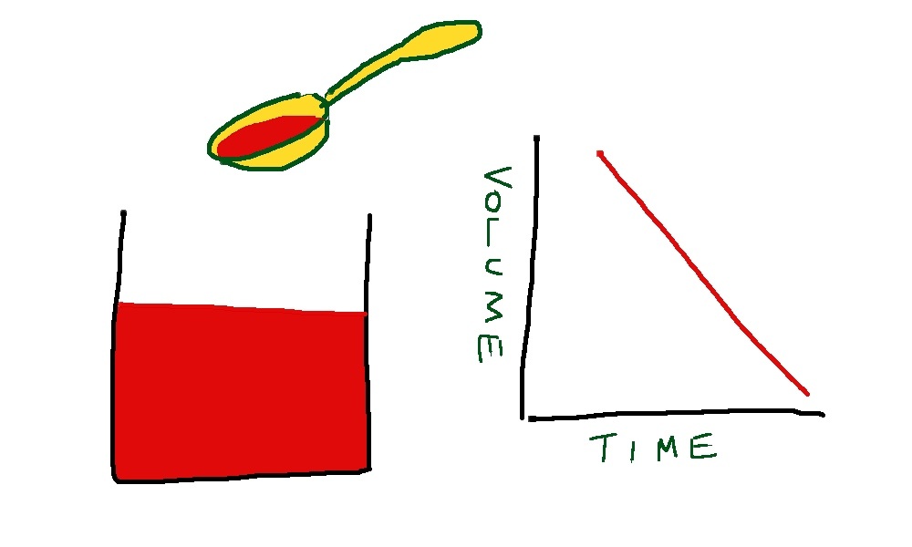
Red blood cells last on average 120 days before they wear out and have to be recycled.  As we have five litres of blood and in each litre there is at least 120 grammes of haemoglobin, it means we have to make, and break down, five grammes a day.  About a teaspoonful.  This all happens inside macrophages in the spleen and liver, as they try to salvage useful parts of the red cells to make new things.

Macrophages are similar to neutrophils but slightly smarter.  Some are good at host defence: they come to the scene after the neutrophils have done their job, do the forensics, and find out what the germs are made of. They then tell the lymphocytes how to make antibodies to protect us in the future.

Other macrophages are good at clearing up the mess, breaking down dead tissue and making things neat and clean.  The spleen and liver macrophages that deal with old red cells are the latter kind. When they take red cells apart, they separate haem from globin, and then take the iron out of the haem.  Next, the porphorin ring is broken to form bilirubin – its molecule of linked pyrrole rings looks like four beach huts in a row.

haem is broken down by the enzyme haem oxygenase to form bilirubin

The bilirubin is then thrown away (I don’t know why it can’t be re-used).  It attaches to albumin and becomes water soluble after conjugating in the liver with a sugary molecule called glucuronate.  The conjugated bilirubin is then transported into the biliary system, stored in the gallbladder, and squirted into the duodenum when we eat fatty food.  In the bowel, it is converted to urobilinogen, which makes poo brown.  Some is reabsorbed and excreted in urine, which makes urine yellow.Slide2

Sylvia had noticed that her poo had changed colour – it had become putty-coloured – and her urine had become much darker. That is because conjugated bilirubin was not getting into her intestines and instead some had leaked into her bloodstream and was appearing in her urine. The whites of her eyes and skin had become yellow because of the very high level of conjugated bilirubin in her blood.

Now to the gallstones.  In Western countries they are most commonly made of cholesterol.  I have already talked about the importance of cholesterol in keeping cell membranes rigid and non-leaky.  Cholesterol is made in large amounts by the liver and transported to other cells gift-wrapped as LDL cholesterol.  It is also used to make bile salts.  It only needs a minor modification of the basic chemical structure of cholesterol to make bile salts – the main component in bile which makes it work – the main function of bile salts is to help us digest dietary fat.

structure of cholesterol

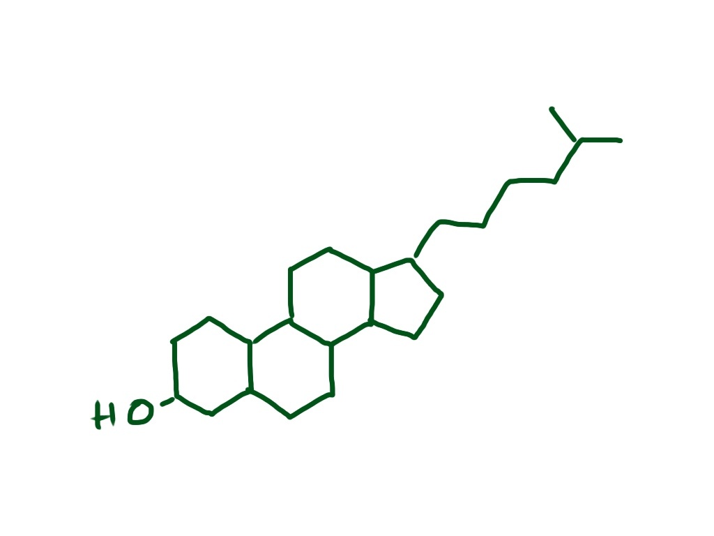
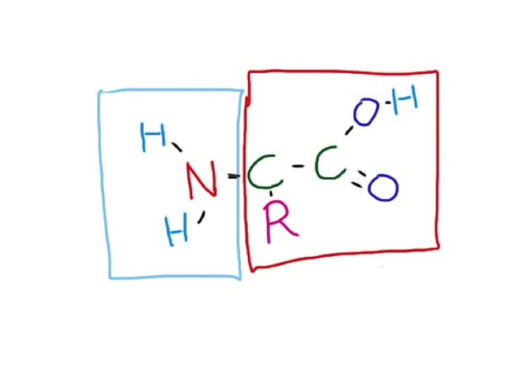
Bile salts are detergents.  Just like washing-up liquid, they emulsify fats, breaking large fat globules into smaller micelles which do not stick to each other, or anything else. Cholesterol is essentially very insoluble in water, or hydrophobic.  When an organic acid group is added – to make bile salts – it gains a hydrophilic or water-attracting group. This is just like soap – a long lipid chain with an organic acid group at the end.  The hydrophobic lipid part is embedded in the tiny fat globule and the hydrophilic groups stick outside in the watery medium of the intestinal contents.

bile acids are a minor modification of cholesterol - hydrophilic OH  and COOH  groups are added to make it into a detergent
bile acids are a minor modification of cholesterol – hydrophilic OH and COOH groups are added to make it into a detergent

As well as bile salts, the liver secretes phospholipids into bile – principally phosphatidylcholine. This, with cholesterol, is the main stuff cell membranes are made of.  It is also an emulsifying agent, and is used extensively in the food industry to keep fats in suspension.

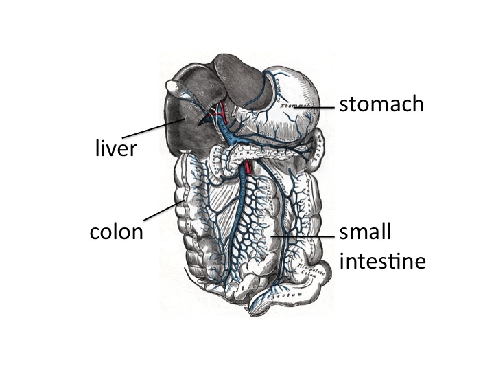
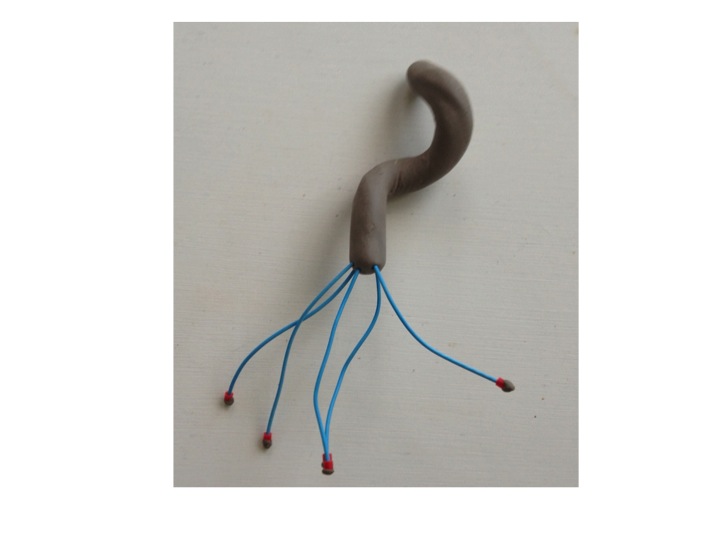
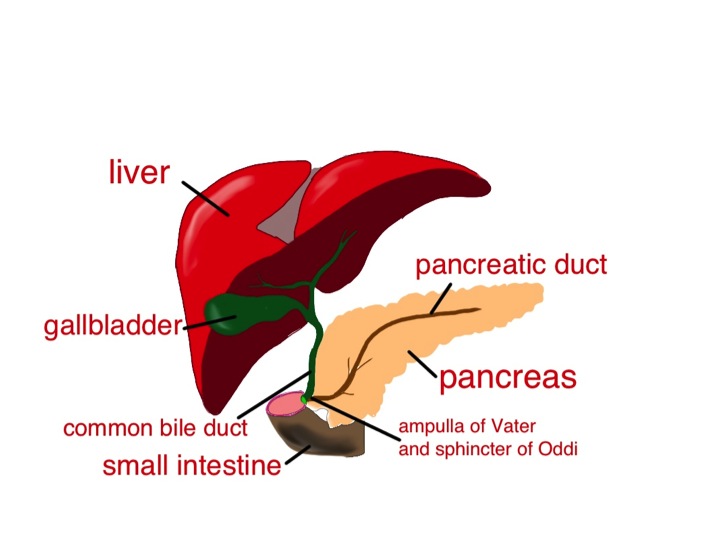
So, when we eat fat, the small intestine detects it and releases the hormone cholecystokinin (CCK).  This causes the gallbladder to contract to send bile into the duodenum, and also makes the pancreas release enzymes, including lipase.  CCK also has some very interesting effects on our brains, making us feel less hungry, and curiously, opposing the effects of opiate drugs.  The bile and pancreatic secretions are both delivered into the duodenum through the same tube – see diagram. The detergent bile salts, emulsifying phospholipid and pancreatic lipase are designed to work together to digest fat.

the hydrophilic parts of cholesterol are all on one side of the molecule - this allow it to act as a detergent - it works well to disperse fat into small globules so that pancreatic lipase can work on it and break it down to fatty acids and monoglyceride
the hydrophilic parts of cholesterol are all on one side of the molecule – this allow it to act as a detergent – it works well to disperse fat into small globules so that pancreatic lipase can work on it and break it down to fatty acids and monoglyceride

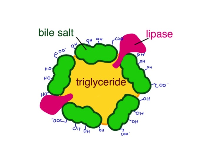
The bile breaks it up into tiny globules and the lipase breaks triglyceride into fatty acids and monoglyceride. These are transported across the intestinal mucosal cell membrane and then the triglyceride is put back together again. Seems a daft system, but it clearly works. The triglyceride is taken away from the intestine not in the portal blood, like most other substances absorbed by the gut, but in the lymphatic system in the form of chylomicrons – small fatty globules covered by a layer of phospholipid. The chylomicrons travel up the lymphatic vessels to emerge into the circulation just below our left clavicle. This means that it avoids passing straight away through the liver. If blood is taken soon after a fatty meal and centrifuged, it will have a milky appearance because of the large amount of chylomicrons.

So why do gallstones form? As well as bile salts, unmodified cholesterol is also secreted into bile. It is only kept soluble by the bile salts and phospholipid – the ratio of cholesterol to bile salts and phospholipid is therefore important. When it goes wrong, cholesterol precipitates out and forms solid stones like those that caused all Sylvia’s recent problems – a bit like when I try to make mayonnaise and it curdles – the fat becomes un-emulsified and separates out.

In some parts of the world, gallstones are made from bile pigment – bilirubin. This can be due to increased red blood cell breakdown resulting from abnormalities such as sickle cell disease, but it’s also common in East Asia, for reasons which are not clear.  A good, comprehensive analysis of why gallstones of all types form can be found at:

http://gastro.ucsd.edu/fellowship/materials/Documents/Gallstones/pathogenesis%20gallstones.pdf

Now the alkaline phosphatase. Bile is alkaline.  Alkaline phosphatase is an enzyme that removes phosphate groups in alkaline conditions.  When bile flow is blocked – by stones, as in Sylvia’s case, or for any other reason – the liver makes much more alkaline phosphatase, some of which appears in the blood.  Bones use a similar alkaline phosphatase to rearrange phosphate groups to make hydroxyapatite – the hard stuff bone is made from.  Most of the alkaline phosphatase normally in our blood is the bone sort.

There is plenty of information about whether alkaline phosphatase is from bone or liver, but very little I can find that suggests why the liver should make this enzyme when the biliary system is blocked.  I think a clue here is that neutrophils also have alkaline phosphatase in their granules, which help kill bacteria.  This is in addition to the myeloperoxidase and esterase mentioned in earlier posts.  Why should a phosphatase be damaging to bacteria?  The answer may well involve techoic acid, an important reinforcing molecule in some gram-positive bacteria.

cartoon of gram positive bacterial cell wall - techoic acid is thought to inhibit the action of lysozyme and protect the peptidoglycan from being broken down - techoic acid contains phosphate groups which may be removed by alkaline phosphatase
cartoon of gram positive bacterial cell wall – techoic acid is thought to inhibit the action of lysozyme and protect the peptidoglycan from being broken down – techoic acid contains phosphate groups which may be removed by alkaline phosphatase

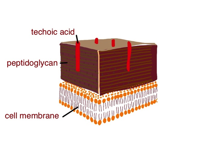
In these microbes, techoic acid strengthens the peptidoglycan cell wall and inhibits the action of lysozyme, yet another enzyme made by neutrophils.  Lysozyme is designed to break some of the sugar-sugar bonds in peptidoglycan – so getting rid of the techoic acid would surely be helpful.  We know that alkaline phosphatase is effective in breaking up techoic acid – see, for instance:

http://jb.asm.org/content/102/3/747.short

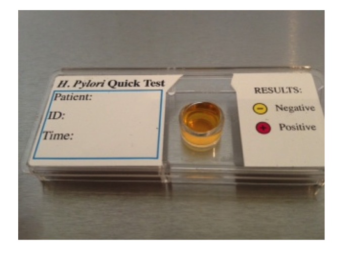
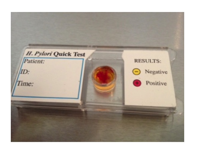
Maybe the liver is producing this anti-bacterial enzyme to help prevent infection when the flow of bile slows down.  Unfortunately, this won’t work with gram negative organisms such as E.coli because they don’t make techoic acid – and it was a gram negative bacterium we grew from Sylvia’s blood culture – the germ that was causing her ascending cholangitis.

Soon after she came in, Sylvia had an ERCP – an endoscopic retrograde cholangio-pancreatoscopy.  The endoscopist managed to remove the impacted gallstone from her ampulla of Vater, and she quickly recovered.  A week later she had her gallbladder removed, including the stones, and now is quite well – she can even eat fish and chips.

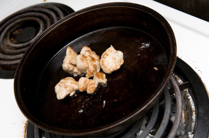
Now to the food link: mayonnaise.

French mayonnaise - made with olive oil and mustard
French mayonnaise – made with olive oil and mustard

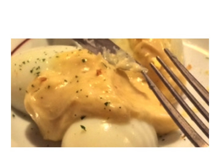
This is an emulsion of lipid, such as olive oil, in an aqueous (watery) medium – vinegar or lemon juice.  The emulsifying agent is raw egg yolk, which – just like bile – contains lots of phospholipid – phophatidylcholine and is also rich in cholesterol.  In France, mustard is also added.  Mustard seed contains mucilage, a gooey stuff that some plants produce made of sugar polymers. (Thanks again to Harold McGee). These also act as emulsifiers and further thicken the mayonnaise, and give it a better taste. When I make cheese sauce, I use a sugar polymer (flour) to achieve a stable emulsion between butter and milk – no doubt the phospholipid in the milk helps too.  It seems likely that in the small intestine, the mucus from the stomach, a similar gloopy sugar polymer, probably has a similar effect to mustard seed mucilage to help with fat emulsification.

vomiting blood and yoghurt

Slide04 More alcohol problems this week. Peter came in vomiting blood yesterday afternoon. Vomiting blood is always a bad thing, but this time it was particularly bad. His long-suffering partner Rita came in with him to tell us what had happened. Peter, aged 69 was too drowsy and confused to tell the story himself. He was a retired barman and had always drunk too much alcohol. He recently went to see the liver doctors because his abdomen had filled up with fluid. They told him that he would die soon if he did not give up drinking. Rita said that he had cut down but was still drinking about 3 pints of strong cider every day. Yesterday lunchtime he was about to sit down to eat when he said he felt very sick. He staggered to the bathroom and promptly vomited what Rita estimated to be a pint of bright-red blood down the toilet pan. Rita called the ambulance. He vomited more blood on the way and by the time they arrived he was pale, sweaty and quite drowsy. She was really worried – she thought about what the liver doctor had said about Peter dying soon if he did not give up drinking – he had not given up.Slide05

When patients with liver disease vomit blood it always makes us worry about bleeding varices. Varices are large, distended veins which appear at the junction between the oesophagus (foodpipe) and stomach in people with liver cirrhosis. Cirrhosis often results from liver damage due to alcohol. Ethanol is metabolised to ethanal (acetaldehyde) and causes damage to liver cells as well as the pancreas (see vodka and sweetbreads below). The liver does have a remarkable capacity to regenerate.

Slide06

I’m not sure if the ancient Greeks knew about liver regeneration when they devised the myth about Prometheus. He made the mistake of giving fire to men, and as a punishment was chained to a rock for eternity. Every morning an eagle would fly down and peck out his liver. During the following day his liver would grow back again, to be pecked out again the following morning. He is still there.

ancient Greek vase showing Prometheus having his liver pecked out by eagle wikimedia common user Bibi Saint-pol
ancient Greek vase showing Prometheus having his liver pecked out by eagle –  wikimedia common user Bibi Saint-pol

Although liver does regenerate when damaged by alcohol, it does so to form nodules of liver tissue with bands of fibrosis in between the nodules. This disturbance of normal architecture impairs blood flow through the liver. As a result the pressure in the portal veins carrying blood from the stomach and intestines to the liver increases. Increase in portal venous pressure results in oesophageal varices. When they burst, rapid death from blood loss is common. Another result of increase in portal pressure is ascites – fluid accumulation in the abdominal cavity – the cause of Peter’s abdominal swelling.

all the blood coming from the stomach and small and large intestine goes into the portal venous system and through the liver to be processed - including removal of ammonia and small amines
all the blood coming from the stomach and small and large intestine goes into the portal venous system and through the liver to be processed – including removal of ammonia and small amines – from Grays anatomy 1918

So he was filled up with a blood transfusion, vitamins (see vodka blog below) and given terlipressin – a drug which constricts oesophageal varices and helps to stop bleeding. He was sent as an emergency to have an upper gastrointestinal endoscopy. In fact he did not have significant varices. He had a bleeding duodenal ulcer. The ulcer was cauterised and injected with adrenaline, a biopsy was taken from his duodenum, and he was sent to the admissions unit.  Part of the duodenal biopsy was put into a CLO test kit.

I talked previously about adrenaline causing muscle tremor and relaxation of bronchial smooth muscle by activating adrenergic beta receptors. It is released by the middle (medulla) of adrenal glands in response to severe stress. The reason Peter was so pale was probably more to do with release of adrenaline than blood loss. This hormone has many other actions to help us survive life-threatening situations. It will also act on beta receptors in muscle blood vessels to increase muscle blood flow – good to get away more quickly from the nasty tiger with dripping fangs that likes to eat humans.

Skin blood vessels have few beta receptors – here adrenaline acts on alpha receptors to cause reduction in blood flow. Similarly, in the lining of the duodenum, adrenaline, when injected by the endoscopist, causes blood vessels to constrict and help stop bleeding by acting on their alpha receptors.

When I ask students why adrenaline reduces skin blood flow they usually say it is to redirect blood to the central circulation where it is more needed. The skin only has about 1% of circulating blood. It is more likely that skin blood flow is reduced to limit blood loss when the tiger’s teeth finally sink into that tasty human flesh.

All this preamble is a good excuse to talk about urea. In our hospital normal blood urea levels are between 3.5 and 6 mmol/l. In the US doctors talk about blood urea nitrogen- the same stuff- normal levels are 20-30mg/l. When Peter visited the liver doctor last month his blood results showed that the concentration of urea in his blood was low – only 1.8 mmol/l (5mg/dl BUN). When he arrived in the ED it was elevated at 14 mmol/l (40mg/dl BUN). His haemoglobin was low at 90g/dl and clotting was deranged with an INR of 2.9. Why was his urea low before and now high? To understand this I need to talk about protein metabolism.

Most of us in the West eat lots of protein. More than we need. In the US and UK adults eat about 100grammes of protein/day, although we only need about 50. If we eat 100grammes of protein a day, we need to get rid of the same amount, unless we are growing, body-building or pregnant. Patients who are ill typically break down more protein than they take in – negative nitrogen balance.

I’ve used arguments about in/out balance for fluid in my previous post and will use it for energy in future posts. Not everything in humans can be understood in terms of in/out balance. For instance, with my 11 year-old son we input high grade educational material and all that comes out is poo and fart jokes.

So what happens to this 100grammes of protein? Protein is a polymer of amino acids. You can make useful plastic out of milk protein – see this youtube video:

http://www.youtube.com/watch?v=pIvAl4lu1uA

Casein also used to be used to make plastic items such as buttons. We now have cheaper and better plastics.

Protein that goes into our mouth is mashed up by our teeth and swallowed. The stomach has the first go at breaking up the protein polymer with the enzyme pepsin, secreted by chief cells in the glands of the stomach. It’s a bit tricky making an enzyme that breaks down protein, because all our cells are made of lots of proteins.  There is the obvious danger that the enzyme will destroy the cell that made it. So pepsin is made in an inactive form – pepsinogen that is only activated when it comes into contact with stomach acid. Then the pancreas has a go. It makes trypsin, carboxypeptidase and chymotrypsin which finish the job, to end up with amino acids. The pancreas has to be pretty careful too, making inactive enzymes which become active one secreted – see:

http://www.physiologymodels.info/digestion/proteins.htm

Amino acids are all of the general formula:

general formula for amino acid - the red box stuff can usually be turned into carbon dioxide and water - the blue box stuff is harder to get rid of
general formula for amino acid – the red box stuff can usually be turned into carbon dioxide and water – the blue box stuff is harder to get rid of

The R is mainly made of carbon, hydrogen and oxygen. We have to dispose of 100g daily. The part in the red box is burned up in mitochondria, and like glucose is turned into carbon dioxide, water and energy – 4Kcal/gramme – 400 Kcalories per 100g. The problem is the bit in the blue box. The NH2 group looks like ammonia, and ammonia is toxic. But don’t worry, we have a way of dealing with this – it’s called the urea cycle. This happens mainly in the liver. I could draw a diagram of the urea cycle, but instead will direct you to a 1 minute youtube video showing how it works:

http://www.youtube.com/watch?v=AoBbVu5rnMs

So ammonia is combined with bicarbonate (or carbon dioxide) and via the urea cycle is made into urea, a very non-toxic substance which is excreted in urine.

two molecules of ammonia combine with one of carbon dioxide to make urea and water - its not as simple as that - but that is what the urea cycle does
two molecules of ammonia combine with one of carbon dioxide to make urea and water – its not as simple as that – but that is what the urea cycle does

The reason Peter’s urea was so low when he saw the liver doctor was because he was drinking too much alcohol and not eating enough protein. Now he has had a sudden high protein meal – blood. Blood contains about 70g/l of albumin and globulins in plasma, and about 130g/l of haemoglobin (mostly protein) in red cells – total 200g/l.  If he has bled a litre of blood from his duodenal ulcer he will suddenly have twice the average daily protein intake in a short time – no wonder his urea level has risen.

But Peter’s liver is not working as well as it should, because he has been drinking too much alcohol for a long time and has cirrhosis. A lot of the ammonia from protein metabolism is not being instantly turned into urea, and instead gets into the systemic circulation. When the brain is exposed to ammonia, it does not function too well. It is not only ammonia, but other short-chain amines which the liver has failed to deal with. Bacteria in the colon chop up amino acids and proteins into all sorts of amine-containing molecules which the liver will normally cope with. Peter’s damaged liver is not able to cope with these substances and they get into his circulation and into his brain. Nobody knows how brains work, but we do know that certain chemicals are important in passing messages from one brain cell to another – neurotransmitters. Many neurotransmitters are small amine-containing molecules, such as catecholamines, serotonin (5-hydroxytryptamine), glutamine and dopamine. It is not surprising that when flooded with a smorgisbord of small amines and ammonia the brain does not work too well. Rita told us that Peter was sleeping all day and awake all night recently – a characteristic feature of hepatic encephalopathy. When we examined him when he came back from endoscopy he had a liver flap, or asterixis. That means when he held out his hands they would independently twitch, with a downward flapping movement- another feature of hepatic encephalopathy which was also making him drowsy. There are lots of examples of liver flap on youtube such as:

http://www.youtube.com/watch?v=Rbv-zaVszlk

The reason his INR was raised was because his liver was not producing enough clotting factors. We gave him a concentrate of clotting factors (octiplex) to correct this, and help stop the bleeding.

It took a couple of hours before his CLO test result was available. A small piece of tissue was removed from his duodenum next to the ulcer. The CLO test refers to Campylobacter Like Organism. Helicobacter pylori is one of these which is a major cause of duodenal ulcers. Helicobacter pylori is an amazing germ. It is a bacterium which can survive in a very hostile environment – the human stomach and duodenum. It is the only germ that can normally survive here. The acidity is fierce, often the pH is down to 1 – really strong hydrochloric acid. There is also a high concentration of nitric oxide and proteolytic enzymes. It doesn’t mind.  Helicobacter refers to the shape – a helix or corkscrew.

model of helicobacter showing corkscrew shape and long flagellae

I talked before about how bacteria find it difficult to swim in mucus. The stomach lining is covered in mucus, but helicobacter’s corkscrew shape helps it swim in this thick and gloopy layer. Staying in the mucus layer helps protect it from the acid and enzymes. Another protection comes from being able to convert urea into alkaline ammonia – keeping the acidity at bay in its immediate environment. The CLO test simply tests the small bit of duodenum for its ability to convert urea into ammonia – it can only do this if has helicobacter organisms in it. Human cells cannot do this. So, in the test plate, there is an indicator, such as phenolphthalein or phenol red which changes colour with alkali.

this CLO test kit contains phenol red - it is yellow when acid and red when alkaline
this CLO test kit contains phenol red – it is yellow when acid and red when alkaline

The helicobacter turns urea into ammonia, making the environment more alkaline which will change the colour of the indicator. Phenol red changes from yellow to red with alkali.

when a piece of duodenum is put into the CLO test vial it will turn red if there are helicobacter pylori organisms - I just used a bit of soap for this photo as I did not have any infected duodenum to hand
when a piece of duodenum is put into the CLO test vial it will turn red if there are helicobacter pylori organisms – I just used a bit of soap for this photo as I did not have any infected duodenum to hand

We gave Peter intravenous omeprazole, a proton pump inhibitor. This stops the parietal cells of the stomach making acid and helps heal the ulcer. We also gave him lactulose. This is a sugar which helps with hepatic encephalopathy. Lactulose is very similar to lactose – the sugar in milk. Lactose is a disaccharide made of the simple sugars glucose and galactose. Lactulose is made of fructose and galactose. I talked earlier (chest pain) about how most of the world’s population have problems drinking milk when they are adult because they have lost the enzyme which breaks down the bond between the two sugars in lactose. No-one has the enzyme to break down lactulose into its two simple sugars. But bacteria do. In the large intestine, lactulose that we have been unable to break down and absorb in the small intestine is turned into lactic acid, which is also then turned into lots of gas, such as methane and hydrogen. Some patients don’t like lactulose because it gives them stomach cramps and makes them fart a lot. But in patients like Peter it is good because the lactic acid reacts with the alkaline ammonia and small amines to inactivate them and reduce their amount in the circulation affecting his brain, making him confused and drowsy. He went home less confused and more or less OK. I hope he stops drinking.

This is where the yoghurt comes in. The food link at last. In yoghurt the milk sugar lactose is broken down by lactobacilli to form lactic acid. This gives it a nice tangy taste that my daughter likes so much she wears the following tee-shirt.Slide08

Vodka and sweetbreads

Drinking too much is becoming a big problem in our admissions unit. We used to see alcohol problems occasionally, now we see them a lot. Today we admitted Kevin, age 44. He had been drinking at least a bottle of vodka every day for the past year or more.

vodka and tonic

His friend called the ambulance because Kevin had terrible upper abdominal pains and vomiting. He didn’t want to come into hospital because he knew he would not get his vital vodka, but relented when he realised that he could not drink anything without throwing up. The problem was that he had acute pancreatitis caused by his drinking.

As soon as he arrived in the emergency department an intravenous cannula was put in and he was given Pabrinex, intravenous fluids and morphine.

pabrinex

Pabrinex is the trade name for a combination of vitamins, mostly  B vitamins, and importantly it contains lots of vitamin B1 or thiamine. It is bright yellow because it contains vitamin B2, also known as riboflavin which is widely used to colour food and drinks such as orange juice (E number 101). If you take too many B vitamin supplements the riboflavin can make your urine a fluorescent bright yellow.  But thiamine is the important one.

We are really keen to make sure that alcoholics get thiamine as soon as they arrive in hospital. Without it they can suffer permanent brain damage.

Kevin is addicted to, and dependent on alcohol. That means he feels very unwell if his blood alcohol levels fall to near zero, so he must keep his intake enough to make sure that does not happen.

Alcohol is removed from the body in a different way from most other substances. Usually the rate at which a chemical such as a drug is removed from the body is dependent on the amount of drug present. To put it another way, the rate of elimination depends on the concentration in the blood. High concentrations mean that every hour a lot is removed, low concentrations much less is removed.

Bucket hole 2

The normal way things work with drug elimination is the bucket-with-a-hole-in-the-bottom method.  When the bucket is full, water gushes out of the hole quickly, but when nearly empty comes out in a trickle – this is called first order metabolism. But alcohol is not handled in this way – if it was it would be a disaster. Alcohol, or ethanol to give its proper chemical name is first turned into ethanal (also known as acetaldehyde) and then ethanoic acid (aka acetic acid). Both ethanal and ethanoic acid are pretty toxic.

Bucket ladle

Ethanol is not metabolised by first-order metabolism, but by zero-order metabolism. The bucket analogy now is to think of someone with a ladle who scoops out a measure of water from the bucket every 10 seconds. The rate at which the bucket empties now is not dependent on the amount in the bucket, but on the size of the ladle. This is a safe way to metabolise ethanol because it limits the amount of ethanal and ethanoic acid which can accumulate – not enough to do serious damage.

Ethanol-2

Have you ever noticed the guy asleep on the floor at the end of the party who has drunk so much that he can’t get himself home? Next time look at his breathing pattern – he will have slightly rapid, deep, and sighing respiration. This is known as Kussmaul breathing and is due to the large amounts of acetic acid being produced from the alcohol he has inadvisedly drunk. (Although I note that the person who has written the Wikipedia article on Kussmaul’s respiration says that this term only applies to those patients about to die from acidosis – not how it is used in most medical wards – perhaps a bit of a pedant?). If the alcohol was being metabolised by a first-order hole-in-the-bucket process he would not survive.

How big is the ladle? – various authorities suggest this is between 10-15 mls of alcohol per hour. If we take the lower figure this equates to 1 unit of alcohol per hour. That means Kevin has to drink 240mls of ethanol every day to keep enough on board to keep his brain happy.  Most vodka in the UK is 40% alcohol by volume. So an average 750ml bottle contains 300mls alcohol – that will do nicely!

The problem is that this 300mls of alcohol has a lot of calories. 300mls is 240 grammes (the specific gravity of ethanol is about 0.8). Each gramme of alcohol provides about 7Kcal so the bottle of vodka has about 1600Kcal of energy. Given that Kevin drinks his vodka with tonic water, which provides 150Kcal/day, and that his requirement to maintain normal weight is 1800Kcal/day suggests that he does not eat many other calories. He admits this.

It reminds me of the Glasgow vegetarian diet – 15 pints of heavy and 2 packets of crisps.

The serious point is that if someone is truly dependent on alcohol, they will be seriously malnourished. Vodka and tonic is not a balanced diet (no, it really isn’t). There are all sorts of nutritional problems that alcoholics encounter, but one really important one is irreversible brain damage due to thiamine deficiency.

What is thiamine, and why does deficiency cause brain damage? Thiamine is vitamin B1, present in many foods, and if you have a varied diet you will not become thiamine deficient. It is important as a co-factor in a number of enzyme reactions, particularly those of the Kreb’s cycle which produce energy from carbohydrates, protein and fats (and alcohol). The brain and heart use more energy than other organs and are therefore more susceptible to thiamine deficiency, causing cerebral beri-beri (Wernicke’s encephalopathy) and wet beri-beri (congestive heart failure).

There is a wonderful, if somewhat disturbing, paper from 1947 by Hugh de Wardener which helped convince the world that thiamine deficiency causes brain damage.

Dr de Wardener was sent to Singapore in 1942, just before the Japanese completely overran the peninsula and captured 80,000 allied troops. They were marched to the notorious Changi POW camp – see

http://www.historylearningsite.co.uk/changi_pow_camp.htm

and

http://www.abc.net.au/changi/life/default.htm

He became a medical officer at Changi, when large numbers of British servicemen were only given small amounts of white rice (with weevils) instead of their usual rations. This, combined with dysentery meant that large numbers succumbed to thiamine deficiency.

thiamine oil

Thiamine keeps the Kreb’s cycle going – you can think of it like a lubricant to keep the wheel turning. When it runs out the Kreb’s cycle comes to a juddering halt. We need about 1mg/day to do this – not much but we absolutely need it. Our bodies can store about 50mg of thiamine, so after about 6 weeks the prisoners of war became ill. They developed the classical signs of Wernicke’s encephalopathy – confusion, nystagmus (wobbly eyeballs), diplopia (double vision) and ataxia (unsteadiness and incoordination). A large number died

Slide1

Professor Hugh De Wardener MBE

Dr de Wardener realised that this was an unparallelled opportunity to study the effects of thiamine deficiency – not something you could ethically do in humans now. He and his colleagues carefully described the clinical features of soldiers suffering from thiamine deficiency, or beri-beri, and when they died, pickled parts of their brains in the small amount of formalin they had available.

Slide0001

The mammilliary bodies are particularly prone to damage by thiamine deficiency. 

When it was clear that the Japanese were in danger of losing the war, the enemy were determined that all records of what had happened at Changi should be destroyed. Hugh de Wardener realised that his precious medical notes were at risk, and he buried them 2-3 feet deep in a recent grave, along with the post-mortem brain specimens, in a 4 gallon tin which was sealed and soldered shut. The tin was later recovered, taken back to England and the paper was written. Pictures of the brain specimens are in the paper. I could not find a free fulltext version on the internet, but suggest you get your library to order it if you can – it is a fascinating read:

De Wardener, H. E. and Lennox, B. (1947) Cerebral beriberi (Wernicke’s Encephalopathy): review of 52 cases in a Singapore prisoner-of-war hospital. Lancet 1, 11 – 17.

I can’t talk about thiamine deficiency and brain damage without mentioning Korsakoff’s psychosis. This is a truly debilitating long-term loss of memory which is vividly described in Oliver Sack’s book “The man who mistook his wife for a hat”. If you have not read this book you need to buy or borrow it, but would warn that you should not plan to do anything important for the following day or two because you will not be able to put it down.

Kevin’s pancreas was damaged by too much alcohol, because ethanol is metabolized to ethanal which damages protein – much the same way that formalin (properly known as methanal) was used by Hugh de Wardener to preserve his brain specimens.

I regularly eat lamb pancreas. It is sold by my butcher as sweetbreads.

Seared lamb sweetbread in a skillet.

Salivary glands, thymus and testicles are also called sweetbreads and taste very much the same as pancreas, and they all look quite similar when viewed under the microscope. Perhaps its not surprising that the three glands which are attacked by the mumps virus are salivary glands, pancreas and testicles. Mumps virus likes them all raw. I’ve never tried testicles but  I like pancreas gently fried in butter on toast with a dribble of balsamic vinegar.