Monthly Archives: October 2013

back pain and blue cheese

These are lambs kidneys - not eaten much in the UK but muched loved in France - rognons de veau
these are lambs kidneys – not eaten much in the UK but much loved in France – rognons de veau

Kirsty was admitted this morning. She is twenty eight  and had been unwell for the last two days. The first thing she noticed was a nasty burning pain when she passed urine. Almost as soon as she had finished peeing she needed to go again. Kirsty went to the doctor yesterday and was prescribed antibiotic tablets. She took one dose and was immediately sick. Then she developed pain in her back, on the left side, under her lower ribs. It started gradually but became almost unbearable. Then she started to feel feverish and shivery. Not ordinary shivery, but uncontrollably shivery – and then she vomited again and again. Her new husband, Sam, drove her up to the emergency department at nine thirty this morning. Their two year old daughter, Ellie, was in the back of the car. In the emergency department Ellie was sitting on Sam’s knee, looking very unconcerned when we talked to Kirsty, who was lying on the trolley.

“I think you have a serious kidney infection” I said, “and we’d better admit you and give you intravenous antibiotics”.

Kirsty was not looking well. She was pale, sweaty, febrile and a bit blotchy with wet hair stuck to her face.  She was clutching her painful back with one hand and holding a vomit bowl with the other. She was happy to come in and be looked after. So was Sam. Ellie did not look so sure.

its really important to take blood cultures before giving antibiotics - otherwise it will be difficult to find out the germs responsible for infection
its really important to take blood cultures before giving antibiotics – otherwise it will be difficult to find out the germs responsible for infection

We took a blood sample and blood cultures, gave her intravenous morphine, paracetamol, gentamicin, co-amoxyclav and cyclizine (an antiemetic). She had pyelonephritis – a bacterial infection of her kidney.

Young women get urinary tract infections much more commonly than young men. It’s to do with anatomy. The female urethra is very short, and germs can quite easily travel up to the bladder and then up the ureters to the kidney. Men get urine infections when they are older and have enlarged prostate glands.

Then they cannot empty their bladders completely and the stagnant urine is more likely to become infected. I was taught that urine is normally sterile in healthy people. It seems this is not the case. All of us have bacteria in our urine in small numbers – if you are interested read: http://www.ncbi.nlm.nih.gov/pmc/articles/PMC3744036/ 

We use urine dipstick testing a lot on the acute medical unit. In theory it should be able to tell us which of our patients have a significant urinary tract infection. In practise it is not quite as useful as it should be. Kirsty’s urine tested positive for nitrites and leukocytes. In young women this is a good test, but we knew she had a urinary tract infection anyway. In elderly women, the tests are often positive even if a serious infection is not evident – perhaps because of the innocent commensal bacteria which are present.

Slide02

How do urine dipsticks work? The test for leukocytes is leukocyte esterase. In infected urine, the leukocytes are polymorphonuclear leukocytes, or neutrophils. I talked earlier (phlegm and horseradish) about how neutrophils, when they get excited by the presence of germs, make the enzyme myeloperoxidase which generates bleach. Esterase is another enzyme neutrophils make which breaks down peptide bonds, and specifically is useful in breaking down the peptidoglycan in bacterial cell walls. I guess its like getting stains out of clothes. Bleach works fine, but the proteolytic enzymes in washing powder can help too.

So what about nitrites? Bacteria like Eschericia coli, which are a common cause of urine infections are known as facultative anaerobes. This means that they can use oxygen to “burn” carbohydrates, protein and fats. But they can also use other “electron acceptors” to do this such as nitrate. Mammalian cells can only use oxygen. When I say mammalian cells, what I mean is mitochondria in mammalian cells – these small structures in our cells are responsible for all the energy generation from glucose, fats and protein. Think what happens when you eat a slice of toast. The amylase in our saliva starts to break down the starch in the toast to form glucose, a process which is finished by amylase in pancreatic secretions. This yields glucose, which is absorbed into the bloodstream. Glucose can be made into energy by the Kreb’s cycle, or citric acid cycle mainly happens in mitochondria. This is a complicated process, but essentially it means that glucose is turned into carbon dioxide and protons and electrons:

The mitochondria don’t get much energy from the Kreb’s cycle, but rely on the protons and electrons produced to make energy by combining them with oxygen. This happens in the electron transport chain:

Bacteria are more versatile than mammalian cells and can get energy out of these protons and electrons, even if oxygen is not available, by using nitrate instead of oxygen:

To do this they need to have the enzyme nitrate reductase. Human cells do not have nitrate reductase, so if nitrate is being turned into nitrite there must be bacteria present. So, if urine contains significant amounts of nitrite, the only way it can get there is if bacteria are using nitrate to “breathe” – a tell-tale sign that infection is present. The reason E.coli is called a facultative anaerobe is that it can survive by making energy if oxygen is present and also when it is not, by using molecules such as nitrate to “breathe”.

our mitochondria can only use oxygen to get energy from electrons and protons derived from glucose - some bacteria have nitrate reductase which can use nitrate as an electron acceptor - nitrite is a by-product- and appears in infected urine as an indicator of infection
our mitochondria can only use oxygen to get energy from electrons and protons derived from glucose – some bacteria have nitrate reductase which can use nitrate as an electron acceptor – nitrite is a by-product- and appears in infected urine as an indicator of infection

When I saw Kirsty later in the afternoon she was much better. The pain had not gone, but was eased by the morphine. Her temperature had come down, and she had stopped vomiting. Her infection was coming under control.

Both gentamicin and co-amoxyclav are very effective in treating urinary tract infections. They are both rapidly excreted by the kidneys and achieve much higher concentrations in the urine than in the bloodstream. Gentamicin has a half-life of about 2 hours in patients with normal renal function. So, lets say we give a person 250mg of gentamicin intravenously. The blood volume is about 5 litres, so the immediate concentration will be 50mg/litre of gentamicin. In two hours about 100ml of urine will be made, and half of the gentamicin previously given intravenously will be in that urine. That is 125mg in 100mls or 1250mg/litre – more than twenty times the concentration in blood. Amoxycillin has a half-life of more like one hour, so achieves even higher urine concentration in comparison to blood.

What do gentamicin and co-amoxyclav do? They are antibiotics that work in quite different ways.

Gentamicin is an aminoglycoside. That means it is a sugar with amine groups. Here is the structure – just three sugars with lots of NH2 groups:

gentamicin is a relatively small molecule with three sugar groups and lots of amine groups (in red) - an aminoglycoside
gentamicin is a relatively small molecule with three sugar groups and lots of amine groups (in red) – an aminoglycoside

It gets into the bacteria and binds strongly to its ribosomes. These are the really important and clever machines in bacteria which make proteins from DNA. Mammalian cells also have ribosomes to make our proteins – but they are not at all the same. They work in the same way but over the past 2 billion years have changed with evolution so they are a different shape and are larger than bacterial ribosomes. Gentamicin does not interfere with mammalian ribosomes. For an antibiotic to be useful it has to damage bacteria but not human cells. Luckily, ribosomes are so different between bacteria and mammalian cells that some chemicals such as gentamicin will selectively bind only to bacterial ribosomes.

mitochondrion - it has all the stuff inside that a bacterium has, but without a tough cell wall
mitochondrion – it has all the stuff inside that a bacterium has, but without a tough cell wall – redrawn from wikipedia – author kevinsong

Other antibiotics such as tetracyclines, macrolides (erythromycin and clarithromycin), chloramphenicol and clindamycin also work by interfering with bacterial ribosomal function. A bacterium with damaged ribosomes has major problems – it cannot make new proteins. That means it cannot divide and make new bacteria. It will be immobilised and suffer a slow and painful death. (Not really, I don’t think bacteria don’t feel pain – but then I don’t have evidence for that). If it is a bacterium which makes a protein toxin, such as staphylococcus causing toxic shock syndrome, turning off protein production with a ribosomal poison such as clindamycin is a good idea – rather than causing bacterial cell wall damage and leakage of more toxin with penicillin therapy.

Gentamicin can cause problems if it is given over prolonged periods, because it can accumulate and cause damage to ears and kidneys. The damage to hearing is probably due to damage to mitochondria. More specifically damage to mitochondrial ribosomes. We only gave Kirsty one dose of gentamicin – problems with this drug usually happen when patients with impaired renal function are given aminoglycosides for several days, or when aminoglycosides are given with other drugs such as vancomycin which can impair renal function.

Mitochondria are thought to derive originally from bacteria. Once upon a time, a long time ago there was a cell that survived well enough by getting energy from glycolysis – turning glucose into pyruvate. This cell did not need any oxygen. It scraped a living producing at most 2 ATP molecules per glucose molecule. Then it had a conversation with a bacterium which said “Hi doll, I could take that pyruvate you make and turn it into another 28 ATP molecules by combining it with oxygen – how about it?” Maybe this conversation happened at the time oxygen had begun to appear in the atmosphere (see great oxygenation event in last week’s blog). “With your looks and my talent we could do Broadway together”. This is technically known as endosymbiosis, where one type of cell engulfs another to work together to their mutual benefit. The result is eukaryotic cells – the cells we are made of. Our cells contain mitochondria that derive from bacteria. They can make lots of energy from glucose and oxygen. The bacteria are looked after and nurtured inside the cells which engulfed them. Like bacteria, these mitochondria have their own ribosomes, that, not surprisingly, are similar to the ribosomes of bacteria that are causing Kirsty’s pyelonephritis. Too much gentamicin can damage mitochondrial ribosomes and cause hearing loss – see:

Click to access 9443888.pdf

We also gave Kirsty co-amoxyclav.

the basic penicillin molecule - the central beta lactam is in the pink circle
the basic penicillin molecule – the central beta lactam is in the pink circle

This is a combination of amoxicillin and clavulanic acid. Amoxycillin is a penicillin. Originally discovered by Alexander Fleming, the original penicillin, benzylpenicillin, has been modified by pharmaceutical companies to be more effective. Unlike benzylpenicillin, amoxycillin is rapidly absorbed by the stomach. It is also effective against gram negative organisms such as E.coli. Penicillins have a beta lactam group. This structure makes it difficult for bacteria to make a vital component of their cell wall – peptidoglycan.  This is a tough polymer made of special sugars and short peptide chains. The beta-lactam group in penicillins is the right shape to get stuck in the cell wall building enzymes and prevent cell walls being made. Our cells do not have cell walls – they just have thin, delicate plasma membranes made of phospholipid and cholesterol. Similarly, although mitochondria are similar to bacteria, they also do not have cell walls. Mammalian cells and their mitochondria are very cosseted and protected in a 5-star luxury apartment with all mod-cons.  They are looked after in a temperature-controlled environment. Oxygen is supplied free and waste carbon dioxide and other unwanted substances taken away continually. Acidity is tightly controlled – pH between 7.35 and 7.45, osmolarity not too high or low. The poor bacterium, in contrast, has to tolerate acid, alkali, high and low osmolarity and a whole host of chemical insults as well as having to find its own food. And then there is the danger of being chased by an angry green neutrophil. No wonder it feels happier with a thick, tough cell wall to protect it. The clavulanic acid is to inactivate beta lactamase – an enzyme some wily bacteria have started making to destroy beta lactam antibiotics. No doubt the bacteria will soon be making betalactamaseinhibitorase enzymes.

There are other ways to selectively attack bacteria without harming human cells. All cells need folate to manufacture nucleic acids. We get our folate from diet – particularly green, leafy vegetables (folate is related to the word foliage). Bacteria do not, in general, have a healthy diet. They instead make the large folate molecule themselves from much smaller molecules. Trimethoprim and sulphonamides prevent bacteria from making folate causing them to suffer and die a “thymineless death” (look it up in Wiki).

Fluoroquinolones such as ciprofloxacin are some of the newer antibiotics which have come into clinical use since I qualified. They were hailed as the new wonder drug, but we now use them relatively rarely because they particularly seem to promote C.difficile infections in frail, elderly people. They work by inhibiting DNA gyrase and Topoisomerase IV. I hope the illustrations will explain how they work:

bacterial DNA is circular
DNA helicase pulls the DNA strands apart so that they can be replicated to make more DNA  when the bacterium divides - but it causes a problem, the DNA becomes supercoiled
DNA helicase pulls the DNA strands apart so that they can be replicated to make more DNA when the bacterium divides – but it causes a problem, the DNA becomes supercoiled
DNA gyrase sorts out this problem - cutting the DNA strand and rejoining it having removed the twist
DNA gyrase sorts out this problem – cutting the DNA strand and rejoining it having removed the twist – fluoroquinolones such as ciprofloxacin stop this enzyme working
having replicated the DNA - the two circular strands are interlinked!
having replicated the DNA – the two circular strands are interlinked!
but topoisomerase IV comes to the rescue and chops the chain and rejoins it to separate the circular strands
but topoisomerase IV comes to the rescue and chops the chain and rejoins it to separate the circular strands- fluoroquinolones also inhibit this enzyme
all sorted
all sorted

The food link this week is blue cheese.
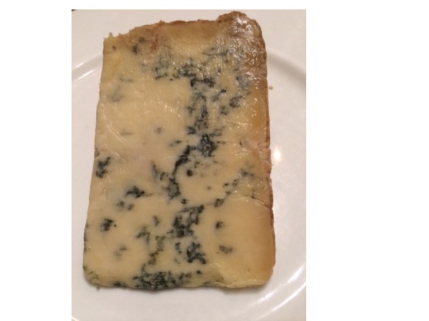
blue stilton
blue stilton – the blue bits are penicillum mould

The reason it is blue is because of the growth of the mould penicillium, which is a grey/greenish blue colour.  The spores are blue, not the fungus itself.
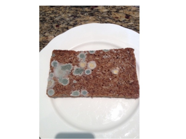
volkornbrot past its sell by date - the blue mould is penicillium - not sure what the yellow stuff is - any ideas anyone?
volkornbrot past its sell by date – the blue mould is penicillium – not sure what the yellow stuff is – any ideas anyone?

The mould in Roquefort and Stilton is P. roquefortii, a close relative of P. notatum (now known as P. chrysogenum), the penicillium mould that Alexander Fleming found inhibiting the growth of staphylococci.

I’ll be in France next week so the next post will be in 2 weeks’ time

vomiting blood and yoghurt

Slide04 More alcohol problems this week. Peter came in vomiting blood yesterday afternoon. Vomiting blood is always a bad thing, but this time it was particularly bad. His long-suffering partner Rita came in with him to tell us what had happened. Peter, aged 69 was too drowsy and confused to tell the story himself. He was a retired barman and had always drunk too much alcohol. He recently went to see the liver doctors because his abdomen had filled up with fluid. They told him that he would die soon if he did not give up drinking. Rita said that he had cut down but was still drinking about 3 pints of strong cider every day. Yesterday lunchtime he was about to sit down to eat when he said he felt very sick. He staggered to the bathroom and promptly vomited what Rita estimated to be a pint of bright-red blood down the toilet pan. Rita called the ambulance. He vomited more blood on the way and by the time they arrived he was pale, sweaty and quite drowsy. She was really worried – she thought about what the liver doctor had said about Peter dying soon if he did not give up drinking – he had not given up.Slide05

When patients with liver disease vomit blood it always makes us worry about bleeding varices. Varices are large, distended veins which appear at the junction between the oesophagus (foodpipe) and stomach in people with liver cirrhosis. Cirrhosis often results from liver damage due to alcohol. Ethanol is metabolised to ethanal (acetaldehyde) and causes damage to liver cells as well as the pancreas (see vodka and sweetbreads below). The liver does have a remarkable capacity to regenerate.

Slide06

I’m not sure if the ancient Greeks knew about liver regeneration when they devised the myth about Prometheus. He made the mistake of giving fire to men, and as a punishment was chained to a rock for eternity. Every morning an eagle would fly down and peck out his liver. During the following day his liver would grow back again, to be pecked out again the following morning. He is still there.

ancient Greek vase showing Prometheus having his liver pecked out by eagle wikimedia common user Bibi Saint-pol
ancient Greek vase showing Prometheus having his liver pecked out by eagle –  wikimedia common user Bibi Saint-pol

Although liver does regenerate when damaged by alcohol, it does so to form nodules of liver tissue with bands of fibrosis in between the nodules. This disturbance of normal architecture impairs blood flow through the liver. As a result the pressure in the portal veins carrying blood from the stomach and intestines to the liver increases. Increase in portal venous pressure results in oesophageal varices. When they burst, rapid death from blood loss is common. Another result of increase in portal pressure is ascites – fluid accumulation in the abdominal cavity – the cause of Peter’s abdominal swelling.

all the blood coming from the stomach and small and large intestine goes into the portal venous system and through the liver to be processed - including removal of ammonia and small amines
all the blood coming from the stomach and small and large intestine goes into the portal venous system and through the liver to be processed – including removal of ammonia and small amines – from Grays anatomy 1918

So he was filled up with a blood transfusion, vitamins (see vodka blog below) and given terlipressin – a drug which constricts oesophageal varices and helps to stop bleeding. He was sent as an emergency to have an upper gastrointestinal endoscopy. In fact he did not have significant varices. He had a bleeding duodenal ulcer. The ulcer was cauterised and injected with adrenaline, a biopsy was taken from his duodenum, and he was sent to the admissions unit.  Part of the duodenal biopsy was put into a CLO test kit.

I talked previously about adrenaline causing muscle tremor and relaxation of bronchial smooth muscle by activating adrenergic beta receptors. It is released by the middle (medulla) of adrenal glands in response to severe stress. The reason Peter was so pale was probably more to do with release of adrenaline than blood loss. This hormone has many other actions to help us survive life-threatening situations. It will also act on beta receptors in muscle blood vessels to increase muscle blood flow – good to get away more quickly from the nasty tiger with dripping fangs that likes to eat humans.

Skin blood vessels have few beta receptors – here adrenaline acts on alpha receptors to cause reduction in blood flow. Similarly, in the lining of the duodenum, adrenaline, when injected by the endoscopist, causes blood vessels to constrict and help stop bleeding by acting on their alpha receptors.

When I ask students why adrenaline reduces skin blood flow they usually say it is to redirect blood to the central circulation where it is more needed. The skin only has about 1% of circulating blood. It is more likely that skin blood flow is reduced to limit blood loss when the tiger’s teeth finally sink into that tasty human flesh.

All this preamble is a good excuse to talk about urea. In our hospital normal blood urea levels are between 3.5 and 6 mmol/l. In the US doctors talk about blood urea nitrogen- the same stuff- normal levels are 20-30mg/l. When Peter visited the liver doctor last month his blood results showed that the concentration of urea in his blood was low – only 1.8 mmol/l (5mg/dl BUN). When he arrived in the ED it was elevated at 14 mmol/l (40mg/dl BUN). His haemoglobin was low at 90g/dl and clotting was deranged with an INR of 2.9. Why was his urea low before and now high? To understand this I need to talk about protein metabolism.

Most of us in the West eat lots of protein. More than we need. In the US and UK adults eat about 100grammes of protein/day, although we only need about 50. If we eat 100grammes of protein a day, we need to get rid of the same amount, unless we are growing, body-building or pregnant. Patients who are ill typically break down more protein than they take in – negative nitrogen balance.

I’ve used arguments about in/out balance for fluid in my previous post and will use it for energy in future posts. Not everything in humans can be understood in terms of in/out balance. For instance, with my 11 year-old son we input high grade educational material and all that comes out is poo and fart jokes.

So what happens to this 100grammes of protein? Protein is a polymer of amino acids. You can make useful plastic out of milk protein – see this youtube video:

http://www.youtube.com/watch?v=pIvAl4lu1uA

Casein also used to be used to make plastic items such as buttons. We now have cheaper and better plastics.

Protein that goes into our mouth is mashed up by our teeth and swallowed. The stomach has the first go at breaking up the protein polymer with the enzyme pepsin, secreted by chief cells in the glands of the stomach. It’s a bit tricky making an enzyme that breaks down protein, because all our cells are made of lots of proteins.  There is the obvious danger that the enzyme will destroy the cell that made it. So pepsin is made in an inactive form – pepsinogen that is only activated when it comes into contact with stomach acid. Then the pancreas has a go. It makes trypsin, carboxypeptidase and chymotrypsin which finish the job, to end up with amino acids. The pancreas has to be pretty careful too, making inactive enzymes which become active one secreted – see:

http://www.physiologymodels.info/digestion/proteins.htm

Amino acids are all of the general formula:

general formula for amino acid - the red box stuff can usually be turned into carbon dioxide and water - the blue box stuff is harder to get rid of
general formula for amino acid – the red box stuff can usually be turned into carbon dioxide and water – the blue box stuff is harder to get rid of

The R is mainly made of carbon, hydrogen and oxygen. We have to dispose of 100g daily. The part in the red box is burned up in mitochondria, and like glucose is turned into carbon dioxide, water and energy – 4Kcal/gramme – 400 Kcalories per 100g. The problem is the bit in the blue box. The NH2 group looks like ammonia, and ammonia is toxic. But don’t worry, we have a way of dealing with this – it’s called the urea cycle. This happens mainly in the liver. I could draw a diagram of the urea cycle, but instead will direct you to a 1 minute youtube video showing how it works:

http://www.youtube.com/watch?v=AoBbVu5rnMs

So ammonia is combined with bicarbonate (or carbon dioxide) and via the urea cycle is made into urea, a very non-toxic substance which is excreted in urine.

two molecules of ammonia combine with one of carbon dioxide to make urea and water - its not as simple as that - but that is what the urea cycle does
two molecules of ammonia combine with one of carbon dioxide to make urea and water – its not as simple as that – but that is what the urea cycle does

The reason Peter’s urea was so low when he saw the liver doctor was because he was drinking too much alcohol and not eating enough protein. Now he has had a sudden high protein meal – blood. Blood contains about 70g/l of albumin and globulins in plasma, and about 130g/l of haemoglobin (mostly protein) in red cells – total 200g/l.  If he has bled a litre of blood from his duodenal ulcer he will suddenly have twice the average daily protein intake in a short time – no wonder his urea level has risen.

But Peter’s liver is not working as well as it should, because he has been drinking too much alcohol for a long time and has cirrhosis. A lot of the ammonia from protein metabolism is not being instantly turned into urea, and instead gets into the systemic circulation. When the brain is exposed to ammonia, it does not function too well. It is not only ammonia, but other short-chain amines which the liver has failed to deal with. Bacteria in the colon chop up amino acids and proteins into all sorts of amine-containing molecules which the liver will normally cope with. Peter’s damaged liver is not able to cope with these substances and they get into his circulation and into his brain. Nobody knows how brains work, but we do know that certain chemicals are important in passing messages from one brain cell to another – neurotransmitters. Many neurotransmitters are small amine-containing molecules, such as catecholamines, serotonin (5-hydroxytryptamine), glutamine and dopamine. It is not surprising that when flooded with a smorgisbord of small amines and ammonia the brain does not work too well. Rita told us that Peter was sleeping all day and awake all night recently – a characteristic feature of hepatic encephalopathy. When we examined him when he came back from endoscopy he had a liver flap, or asterixis. That means when he held out his hands they would independently twitch, with a downward flapping movement- another feature of hepatic encephalopathy which was also making him drowsy. There are lots of examples of liver flap on youtube such as:

http://www.youtube.com/watch?v=Rbv-zaVszlk

The reason his INR was raised was because his liver was not producing enough clotting factors. We gave him a concentrate of clotting factors (octiplex) to correct this, and help stop the bleeding.

It took a couple of hours before his CLO test result was available. A small piece of tissue was removed from his duodenum next to the ulcer. The CLO test refers to Campylobacter Like Organism. Helicobacter pylori is one of these which is a major cause of duodenal ulcers. Helicobacter pylori is an amazing germ. It is a bacterium which can survive in a very hostile environment – the human stomach and duodenum. It is the only germ that can normally survive here. The acidity is fierce, often the pH is down to 1 – really strong hydrochloric acid. There is also a high concentration of nitric oxide and proteolytic enzymes. It doesn’t mind.  Helicobacter refers to the shape – a helix or corkscrew.

model of helicobacter showing corkscrew shape and long flagellae

I talked before about how bacteria find it difficult to swim in mucus. The stomach lining is covered in mucus, but helicobacter’s corkscrew shape helps it swim in this thick and gloopy layer. Staying in the mucus layer helps protect it from the acid and enzymes. Another protection comes from being able to convert urea into alkaline ammonia – keeping the acidity at bay in its immediate environment. The CLO test simply tests the small bit of duodenum for its ability to convert urea into ammonia – it can only do this if has helicobacter organisms in it. Human cells cannot do this. So, in the test plate, there is an indicator, such as phenolphthalein or phenol red which changes colour with alkali.

this CLO test kit contains phenol red - it is yellow when acid and red when alkaline
this CLO test kit contains phenol red – it is yellow when acid and red when alkaline

The helicobacter turns urea into ammonia, making the environment more alkaline which will change the colour of the indicator. Phenol red changes from yellow to red with alkali.

when a piece of duodenum is put into the CLO test vial it will turn red if there are helicobacter pylori organisms - I just used a bit of soap for this photo as I did not have any infected duodenum to hand
when a piece of duodenum is put into the CLO test vial it will turn red if there are helicobacter pylori organisms – I just used a bit of soap for this photo as I did not have any infected duodenum to hand

We gave Peter intravenous omeprazole, a proton pump inhibitor. This stops the parietal cells of the stomach making acid and helps heal the ulcer. We also gave him lactulose. This is a sugar which helps with hepatic encephalopathy. Lactulose is very similar to lactose – the sugar in milk. Lactose is a disaccharide made of the simple sugars glucose and galactose. Lactulose is made of fructose and galactose. I talked earlier (chest pain) about how most of the world’s population have problems drinking milk when they are adult because they have lost the enzyme which breaks down the bond between the two sugars in lactose. No-one has the enzyme to break down lactulose into its two simple sugars. But bacteria do. In the large intestine, lactulose that we have been unable to break down and absorb in the small intestine is turned into lactic acid, which is also then turned into lots of gas, such as methane and hydrogen. Some patients don’t like lactulose because it gives them stomach cramps and makes them fart a lot. But in patients like Peter it is good because the lactic acid reacts with the alkaline ammonia and small amines to inactivate them and reduce their amount in the circulation affecting his brain, making him confused and drowsy. He went home less confused and more or less OK. I hope he stops drinking.

This is where the yoghurt comes in. The food link at last. In yoghurt the milk sugar lactose is broken down by lactobacilli to form lactic acid. This gives it a nice tangy taste that my daughter likes so much she wears the following tee-shirt.Slide08

heart failure and haggis

Heart failure is a very common reason to be admitted to our hospital. John, age 73, had a very typical story. He was well until about 3 years ago when out of the blue he had a major heart attack. Life has never been quite the same since. He has to take lots of tablets – aspirin, statins, beta blockers, ACE inhibitors and several fish oil capsules.

His wife worries about John a lot. She no longer makes him steak and kidney puddings and has stopped him smoking cigars when he goes out with his friends on a Saturday night. He has bought a dog and now takes much more exercise and drinks a lot less beer. But life wasn’t too bad until he noticed he was getting more short of breath when he took the dog for a walk a few weeks ago. Then he noticed ankles were swelling – his socks would leave deep rings around his ankles which disappeared in the morning. For the past week he found he could not lie flat without feeling short of breath.

Then the night he was brought into hospital he woke up gasping for breath. He was pale and sweaty and his wife got really worried and called the ambulance. By the time the paramedics got there he was still quite breathless and his ECG was abnormal, so they brought him in to hospital. He was given some nitroglycerin spray under his tongue- this helped a lot. In the emergency department he was given a dose of intravenous furosemide that made him “pee for Britain”.

We examined him on the admissions unit and found he still had some swelling in both his ankles which left dents when squeezed – bilateral pitting oedema- what was once known as cardiac dropsy. His neck veins were fuller than normal and he still had crackles at the base of his lungs when we listened. He had pulmonary oedema due to heart failure.

I really don’t like the term heart failure. You will know that I’m not one for using long Latin words but something like “impaired cardiac function” might be a less alarming label.

So why did John’s ankles swell up and why did he become breathless? When I ask students and junior doctors they usually say something about back pressure and right heart failure. Maybe it’s just in the UK, but I suspect there is a lot of confusion and misunderstanding about the physiology of heart failure. Pulmonary oedema is about back-pressure, but raised central venous pressure and peripheral oedema is not. So what causes ankle swelling in heart failure? What follows is my understanding of what is a complicated problem. I’ll start slowly and it may take a while to get to the answer. This is the most difficult thing I’ve tried to explain so far.

First I want you to imagine a simple pump. The pump is a hollow rubbery container which works because it is made of muscle which contracts rhythmically. At each contraction the volume in the chamber is much less than when it is relaxed. For a normal left ventricle about 60% of the blood is squeezed out each time it contracts (ejection fraction).

the ventricles are made of muscle which squeezes fluid - without valves it would not pump in one direction
the ventricles are made of muscle which squeezes fluid – without valves the heart would not pump in one direction

To make the blood go only in one direction, it is important to have valves at the inlet and outlet of the pump. There are three things helping to fill the pump when the muscle is relaxed. There needs to be a filling pressure, represented in the diagram below by the height of fluid in the left-hand reservoir. In the left side of the heart this is pulmonary venous pressure, in the right it is central venous pressure, most easily measured by looking at the height of blood in the neck veins. But lets first of all think about a simpler system with only one pump and one filling reservoir. Many years ago Frank and Starling discovered that if the filling pressure of a heart chamber affected how well it pumped blood out. The higher the filling pressure the stronger the contraction, making the pump work harder.

the higher the column of blood in the reservoir, the harder the heart pumps - it now has valves so the blood can only go in one direction
the higher the column of blood in the reservoir, the harder the heart pumps – it now has valves so the blood can only go in one direction

A lot of hollow things in our body work in a similar way. The uterus starts to contract when it gets stretched enough by the increasing size of the unborn baby. Our intestines work much harder if they are blocked, causing the wall to be stretched – this results in colic. The same is true for the gallbladder and ureters- both painfully contract when a stone causes a blockage and the muscular wall is stretched.

The second factor helping our heart chambers to fill up is suction. A turkey baster bulb will suck up fluid because the rubbery stuff it is made of wants to spring back to its normal shape. The same is true for the ventricles of the heart – they actively suck fluid in while relaxing during diastole. I am told that if you put an isolated animal heart into a bucket of oxygenated physiological saline solution it will move around like a squid – sucking liquid in and squirting it out to propel it along in the fluid. It could only do that if the ventricle actively sucks in fluid.

The third important factor in ventricular filling is the contraction of the atria. These give a bit of extra stretch to the ventricles by pumping in some more blood  to help encourage them to pump it out a bit harder.

The heart pumps blood around a circuit – the circulation. It first goes into the aorta, but the vessels get progressively smaller in diameter, ending up with capillaries which have an internal diameter of less than that of a red blood cell – about 7 microns.

capillaries have just one layer of cells and are just big enough to allow a red blood cell through

Pushing blood through the large vessels is very easy, but much more difficult through the smaller vessels. There is a strange relationship between the amount of resistance to flow and the internal diameter of a pipe which was discovered by Pouseuille- resistance is inversely proportional to the fourth power of the internal diameter. In practice this means that most of the resistance to flow is in small vessels with a hole down the middle measuring  less than one tenth of a millimetre – these are also known as resistance vessels. Because of Pouseuille’s law, resistance vessels can constrict or relax a very small amount to cause a large change in flow to any particular organ – depending on its requirements for oxygen.

Back to the circuit. The arteries are thick-walled, with a relatively small hole down the middle. Veins are thin walled and can accommodate more volume. So most of the blood in our circulation is in the veins. Blood is pumped into the large arteries, is squeezed through the resistance vessels, and then ends up in the veins. You will not be surprised to know that the amount of pressure in the large arteries depends both on how hard the pump is pumping – cardiac output, – and how much resistance to flow there is in the small arteries – peripheral vascular resistance. Pressure = Flow x Resistance.

One thing our bodies really care about is the pressure in our large arteries – commonly known as blood pressure. It is continually monitored by pressure-sensing devices in the aorta and carotid arteries. These baroreceptors send messages to the brain stem (medulla). If the pressure is low, the medulla sends messages via the sympathetic nervous system to the heart to make it pump harder, and to the resistance vessels to make them constrict a bit to get the pressure back to normal. The medulla also sends messages to the veins to make them constrict – we will see why in a moment.

Looking at our simple circuit, blood pressure is determined by how hard the heart is pumping, and how much resistance to flow there is in small vessels. What determines the pressure on the venous side – the filling pressure of the reservoir (on the left side in the diagram)?

blood moves around in a circuit - the pressure (height of column of blood) in the reservoir will not change if the pump is pumping fast or slowly - but the pressure on the arterial side will very much depend on how much the pump is pumping
blood moves around in a circuit – the pressure (height of column of blood, or central venous pressue) in the reservoir will not change if the pump is pumping fast or slowly – but the pressure on the arterial side will very much depend on how much the pump is pumping

If the heart pumps less hard, or the resistance changes – I hope you can see that neither of these things will make a difference to filling pressure in the reservoir in this simple system. When students talk about “back pressure” they seem to forget that the blood goes round in a circuit. Failure of any part of the heart will not, on its own, affect filling pressure of the right side of the heart. Central venous pressure is determined by only two things: the amount of fluid in the system and how much veins are constricted. The amount of fluid in the circulation is affected by (a) how much fluid we drink every day and (b) how much we lose. Most of the fluid loss is in the form of urine (see previous post). It is the job of our kidneys to regulate the volume of blood in our circulation. Normally they do a fabulous job. If I drink a pint of beer, half an hour later I will be needing to have a pee – lots of dilute urine has been produced to get rid of all the extra water. If I eat two packets of crisps with the beer, over the next few hours my kidneys will get rid of the extra salt with no problem. Kidneys work in a timescale of hours.

central venous pressure is very much affected by the volume of blood in the circulation, which in turn depends on the balance between fluid input and output - cups of tea and daily urine volume
central venous pressure is very much affected by the volume of blood in the circulation, which in turn depends on the balance between fluid input and output – cups of tea and daily urine volume

Venous constriction works in a timescale of seconds. If I am in a chair and about to get up and walk around, my brain stem will send messages via the sympathetic nervous system to my veins to tell them to constrict. Squeezing this complex network of floppy tubes in my legs, belly and chest will increase cardiac filling pressure and, as Frank and Starling discovered, increase the force of contraction of my heart to supply my muscles with more oxygen. Understanding of how heart failure causes problems needs an understanding of how kidneys and veins work, not just the heart.

Now I am going to make the circulation model a bit more complicated. We have two pumps, not one. They are joined in series. The right heart pumps blood into the lungs. It returns to the left heart which pumps it round the rest of the body. The diagram shows the two pumps separated, but of course in real life they are part of the same organ.

this diagram shows the two sides of the heart separated - the pressure in the pulmonary vein is higher than the central venous pressure on the right side
this diagram shows the two sides of the heart separated – the pressure in the pulmonary vein is higher than the central venous pressure on the right side

The fact that they are arranged in series clearly could lead to problems. What if the right heart tries to pump more than the left? Why is this not normally a problem? The way the system is set up is that the right heart pumps blood through the lungs into the pulmonary veins. Here the pressure is higher than the normal 7cm water central venous pressure – in health about 16cm water. This higher pressure in the pulmonary vein is useful priming device so the left ventricle, which is bigger and stronger, can be rapidly filled and has plenty of pressure to stretch the rubbery myocardium to enable it to contract hard and generate adequate systemic arterial blood pressure. When right heart output increases, the pulmonary venous pressure will temporarily increase, and left ventricular output will in turn increase because of the Frank-Starling law, and subsequent reduction of pulmonary venous pressure to normal. The end result is that the output of the left side of the heart matches that of the right, and pulmonary venous pressure has not changed much – all sorted. This does mean, if you think about it, that the cardiac output is being controlled by the right heart – it pumps what it wants and the left heart has to follow suit. It is not always the case that the bigger, stronger partner is in control.  John has found that out recently.

What controls right heart output? Well the brain stem has a big say, by constricting veins and increasing filling pressure and by sending messages to the heart via sympathetic nerves. But the kidneys are also really important – by altering fluid balance they can control filling pressure. Kidneys are involved in turning the dials and can really mess things up when they get it wrong, as we shall see.

Lets get back to John. His left ventricle was damaged by a sizeable myocardial infarction a few years ago (see chest pain and horsemeat lasagne below). Soon after had an echocardiogram which showed that the part of his left ventricle was not contracting as well as it should – the overall ventricular ejection fraction was found to be 44%.  It was probably about 60% before. The rest of his left ventricle which was not damaged has to work harder to maintain his cardiac output and blood pressure. Heart muscle is different from ordinary skeletal muscle – the sort which makes our arms and legs work. Skeletal muscle responds very well to being worked hard. It grows bigger and stronger to more exercise we take. The heart muscle does this to an extent, but eventually it seems to give up and stop working so well. I don’t think anyone quite understands why this is. If experimental animals are infused with isoprenaline, a drug which makes the heart beat strongly by stimulating heart muscle cells in a similar way to noradrenaline released by sympathetic nerves, their hearts will fail in about 2 weeks. Beta blockers stop this sympathetic stimulation, and patients with heart failure live longer if they take them regularly.

Poor cardiac output stimulates the kidneys to produce renin. This is an enzyme which generates angiotensin I. Conversion of angiotensin I to angiotensin II on the surface of blood vessels helps restore blood pressure but also has a bad effect on heart muscle. It seems to make the muscle cells produce damaging free-radicals and oxidising agents which cause cardiac muscle death. ACE inhibitors prevent the formation of angiotensin II and protect the heart. But despite these drugs, once the ventricle is badly damaged, often the remaining heart muscle starts to fail after a period of time. Clearly in John’s case, another small myocardial infarction may have tipped him into symptomatic heart failure, despite the statins, omega-3 (fish oil), aspirin, and lack of steak and kidney pudding.

What happens when the left ventricle fails to pump properly? The right heart output is determined by central filling pressure and sympathetic activity. If it pumps blood through the lungs to the pulmonary veins, and if the left ventricle is not working, it will cause the pulmonary venous pressure to rise – lets say from the normal 12cm water to 20cm water. This rise in pressure will force the damaged left ventricle to pump harder until it can remove all the blood from the lungs that is being delivered. The right heart will not be too pleased with this- it is trying to pump what it thinks is the right amount of blood but is having a problem. As the pulmonary venous pressure rises, assuming the resistance to flow through the lungs does not change, the pressure in the pulmonary artery rises by the same amount as the rise in pulmonary venous pressure. The right ventricle will have to do more work to pump out blood into the lungs. Now, the left heart normally does not mind pumping at high pressure and doing lots of work – that’s what it is designed for. The right heart is just no good at it – it’s not lazy but it just does not have the muscle. (I put that bit in in case my wife decides to read this).

when the left ventricle fails it cannot pump blood out of the lungs and pulmonary venous pressure rises - reduced arterial pressure is sensed by baroreceptors and the kidney - reflex venoconstriction and reduction in urine output lead to a rise in central venous pressure which makes the right heart pump more blood into the lungs - increasing pulmonary oedema - fluid retention by the kidneys is the cause of peripheral oedema, not "back pressure"
when the left ventricle fails it cannot pump blood out of the lungs and pulmonary venous pressure rises – reduced arterial pressure is sensed by baroreceptors and the kidney – reflex venoconstriction and reduction in urine output lead to a rise in central venous pressure which makes the right heart pump more blood into the lungs – increasing pulmonary oedema – fluid retention by the kidneys is the cause of peripheral oedema, not “back pressure”

So, as a result the right heart can pump out less blood and we are left with a new balance – lower cardiac output and higher pulmonary venous pressure. The right heart is under strain because it is having to pump against higher pressure and the left heart is under strain because its filling pressure is higher than normal, making the undamaged parts also work harder than normal.

That’s fine until the kidneys and the brain get involved. They were doing a good job when John was healthy but in a crisis they make some bad management decisions. When cardiac output drops, blood pressure drops in proportion. Baroreceptors sense this and let the medulla know “Houston, we have a problem”. Increased sympathetic supply to small blood vessels is a good idea. Noradrenaline is released from nerves on the surface of blood vessels acting on alpha 1 receptors in the smooth muscle cell membrane. This causes the vessel to contract, making the holes down the middle smaller. Peripheral resistance increases and so does blood pressure.  That was a good decision. There are similar sympathetic nerves which supply the heart. They are activated and the nerves again release noradrenaline, this time acting on beta receptors. The effect is to increase the rate and force of contraction of the heart, increasing cardiac output and blood pressure. Fine in theory, but this increase in work rate will increase cardiac oxygen consumption. John’s heart has a problem with getting enough oxygen because his coronary arteries have been ruined by too many steak and kidney puddings. Sympathetic stimulation can do more harm than good –that’s when reading the theory book goes wrong – a bad decision. And that is why we us beta blockers in patients with coronary artery disease and give them in patients who have had a myocardial infarction.

Another bad decision is to send messages to the veins to constrict. Increasing right heart filling pressure seems like a good idea. The right heart responds by pumping more blood into the lungs, not aware that the left heart is having a problem. If the pulmonary venous pressure goes above 25cm water there are serious problems. Fluid starts leaking out of the circulation in the lungs and causes pulmonary oedema. The fluid causes swelling of the gas-exchange membrane which allows oxygen to pass from the lungs to the blood, and for carbon dioxide to get out. Increased carbon dioxide concentration and reduced oxygen in the bloodstream are sensed by chemoreceptors in the carotid artery and the medulla (the respiratory centre which controls breathing is a close neighbour to the vasomotor centre which controls blood pressure). Houston panics. John (and his wife) also panics – he is fighting for breath. Houston is in panic mode – not only are its baroreceptors and chemoreceptors telling it that things are going wrong – messages are coming down from the White House – the cerebral cortex.

“More sympathetic stimulation” says Houston. Another bad decision. The increased sympathetic increases venous constriction and right heart filling. We are in a bad visious cycle, throwing petrol onto the flames. Just when Houston was beginning to give up Superwoman arrives. The nice ambulance woman has been trained not to panic – she’s seen it all before. It took her about 2 seconds to realise John had acute pulmonary oedema. She sat him up, gave him oxygen and got him to open his mouth and sprayed nitroglycerin under his tongue. Don’t worry dear- you’ll be fine in a moment. And he was. Reassurance works wonders in acute pulmonary oedema by reducing panic. Nitroglycerin or glyceryl trinitrate as we call it in the UK also works very well by selectively dilating veins, having less effect on the small resistance arteries. This reverses the bad vicious cycle, reducing right heart filling pressure, reducing right ventricular output and reducing pulmonary oedema, which reduces carbon dioxide levels in the blood which makes Houston much happier “was that a bird or a plane?”

I know of no evidence to support this, but I feel pretty sure the reason opiates like morphine work to reduce pulmonary oedema is to supress the respiratory centre and reduce central sympathetic output. Opiates certainly don’t work directly on veins to cause vasodilatation.

The problem with the medulla and normal cardiovascular reflexes is that they were designed to get us out of trouble when blood pressure falls from volume loss from trauma, diarrhoea or sepsis. Blood pressure drop from left ventricular failure is not in the manual. Primitive humans did not eat steak and kidney puddings.

The kidney is no better when it comes to heart failure. It knows there is a problem with cardiac output because it can sense it not getting enough blood. It makes the wrong assumption that this is due to circulating volume loss. The kidney’s response to low cardiac output is to stop producing urine. John is still drinking cups of tea and eating salty food, but he is not excreting it. The salt and water has to go somewhere – it increases the volume of blood in the circulation and increases right heart output, predisposing towards pulmonary oedema. The increase in blood volume can only go so far before it leaks out of the circulation and then equilibrates with extra-vascular interstitial fluid to produce ankle oedema. The only way to sort this out is to give diuretics like furosemide. This will force the reluctant kidneys to produce more urine and get the input/output balance right.

Many patients are fearful of taking daily furosemide, thinking that it will make them pass lots of urine all the time and make their lives difficult. Certainly, after the first few doses, daily urine volume increases, but it does not take much thinking about to realise that urine volume will soon settle down to be the same as the volume of fluid drunk each day. It can alter the pattern of urine output, so that for six hours after taking furosemide the volume is higher than normal, but for the next eighteen hours it is less. It might mean that patients need to get up at night to pass urine less often. Furosemide used to be called Lasix, because its effect lasts six hours.

I have explained why patients with heart failure benefit from nitroglycerin, morphine, beta blockers and furosemide. What about ACE inhibitors. As well as producing of free radicals in the heart, angiotensin II has an effect on veins. It does not directly constrict them, but enhances the effect of sympathetic activation originating in the medulla. That means that if the circulating levels of angiotensin II are very high, which is the case in heart failure, normal exercise will result in an exaggerated constriction of veins, an excessive rise in central venous filling pressure and too much blood being pumped into the lungs which the damaged left ventricle can’t handle. This will result in breathlessness. The main benefit patients with heart failure notice when they start ACE inhibitors is a reduction of breathlessness on exertion. The reduction of free radical production is a separate long-term benefit which will keep John alive longer.

So why do students (and doctors) talk about right heart failure as the cause of fluid retention and raised venous pressure when the damage is to the left side of the heart? I think the most likely reason is that in patients with true right heart failure, secondary to severe lung disease or pericardial tamponade, oedema can become really impressive. But again, the cause is not “back pressure”. If the right heart fails to pump properly, say the cardiac output drops from 5 litres/min to 2 litres/min, then the left heart can only pump out 2 litres/min. This means that the kidney again mistakenly goes into shutdown mode and fluid accumulates. The difference here is that pulmonary oedema is not going to be a problem, as the left heart is working normally, keeping pulmonary venous pressure normal. Under these circumstances patients sometimes present with gross oedema in their legs and fluid in their abdominal cavity, without presenting earlier with breathlessness. Thus historically, gross oedema is associated with right heart failure.  Unless you can think of a better reason.

I can’t finish discussion of heart failure without mentioning preload and afterload. Anyone who mentions these terms when talking about humans, in general, is using the terms inappropriately. Preload and afterload were terms invented, I think, by Braunwald and Epstein. They were two brilliant cardiological physiologists who were interested in how filling pressure and arterial pressure affected cardiac ventricular function. They devised very clever experiments on isolated rat heart ventricular strips in organ baths. They wanted to look at the effect of increased filling pressure – so they put a spring in the organ bath to produce more tension and looked at how that affected contraction – they called it preload but never meant it to be anything but a proxy for filling pressure. Similarly, they thought “how can we model how the ventricle contracts against higher arterial pressure?”  They cleverly added weights to be pulled up by the contracting ventricular strip – to make the ventricle do work. They called this afterload, but again did not mean it to be used to describe the in-vivo situation.  If I ask one of the junior doctors “what is that patient’s preload?”  there is no answer. They can tell me that the neck veins are distended. If I ask “what is that patient’s afterload?” they will rightly look at me strangely. But of course they can tell me what his blood pressure is.

haggis

The food link this week is haggis. This is traditionally made from heart, lungs and kidney. Particularly appropriate for this discussion on heart failure. I like haggis roasted, not boiled. When roasted the skin, made from a sheep’s stomach, goes all brown and crispy. You must serve it with neeps (bashed swede, or rutabaga in the US) and tatties (mashed potato). Gravy is good but not necessary if you have a glass of whisky with it. Unfortunately I do suspect it is not much better than steak and kidney pudding for my coronary arteries as it also contains suet – the most saturated fat available. But it does have oatmeal.

Broken arm and salami

The NHS is having a bit of a crisis in the UK, struggling to look after an increasingly frail, elderly population. This week we admitted Agnes. When I saw her name in the casenotes I knew she was going to be elderly. Some names popular in the 1920s have become fashionable again, such as Daisy, Sophie and Amy. Not so for Agnes. She is 93 and was just about managing at home on her own. She has diet-controlled diabetes and recently had a hip replacement.

She slipped when getting out of the bath and fell on her outstretched hand, breaking her right wrist. The alarm she normally keeps around her neck was on the shelf over the bathroom sink. She did not have the strength to get up and nobody heard her calls for help. Agnes lay on the floor for more than a day without food or water.  Eventually her three-times-a-week carer called and found her and called the ambulance.

typical Colle's fracture of the wrist - from Wikimedia commons by
typical Colle’s fracture of the wrist – from Wikimedia commons by Ashish j28

She was already a bit happier when she arrived in the emergency deparment after she had been given some fluids and pain relief by the ambulance crew. She was very sore down one side of her body with obvious large bruises over her buttocks, thighs and arms. The emergency department doctors and nurses had fixed her wrist and put it in a plaster.

Her blood results were worrying. Her creatinine level was raised to over double the normal value for her age, and much higher than it had been after her hip operation 3 months ago. Her creatine kinase level was over 20,000, the normal being below 150.

We were worried because muscle damage can cause severe kidney damage. We knew she had a lot of muscle damage because her creatine kinase level was so raised. Creatine kinase is an enzyme which is vital for normal muscle function. Kinase means an enzyme which adds a phosphate group to creatine. Creatine and creatine phosphate are small molecules which are found in large amounts in muscle – what does creatine do?

Muscles need energy to work. This is supplied by mitochondria which use glucose and other energy sources such as fats and protein. If you throw sugar or fat on a fire it will burn rapidly and provide heat. Mitochondria “burn” energy sources by eventually combining them with oxygen in a much more controlled way. Instead of producing heat, the energy is converted into a chemical bond. Adenosine di-phosphate (ADP) is converted into ATP – adenosine triphosphate. Addition of this extra phosphate is rather like pulling the bowstring on a crossbow. The energy is ready to use to fire the bolt when needed.

The energy stored in ATP is in the chemical bond of the third phosphate group. This energy-containing ATP can then be used by the cell to make muscles contract or perform a whole host of housekeeping functions such as making cholesterol (see previous post).  Muscle needs lots of ATP, so muscle cells have lots of mitochondria. The problem is that ATP does not store well. That’s where creatine phosphate comes in handy. It stores well and can rapidly regenerate ATP from ADP.

creatine and creatinine are small molecules which are in chemical equilibrium

Creatine phosphate is sold in health food shops. There is some evidence eating it can improve exercise performance by increasing creatine in muscles. Creatine is spontaneously converted to a similar molecule, creatinine. This is produced in similar amounts in all of us every day, depending how much muscle we have got, and excreted unchanged by the kidneys.

So why is the small molecule creatinine excreted in kidneys, but molecules we want to keep are not? Kidneys work in a strange way. When we want to get rid of unwanted rubbish in our houses we find the things we don’t want and put them in the bin and take them out to be collected by the waste disposal team. Kidneys do it differently. They do the equivalent of taking all the contents of our house (not large things like furniture) and putting them in the garden. Then they find all the things they want to keep – such as water, sodium, potassium, small proteins and hydrogen ions, and take them back in the house.

pigs kidney
pigs kidney

Before she fell over, Agnes’s kidneys were filtering about 80mls/min through her kidneys. We can calculate this from knowing her age, sex and creatinine level using a modification of a really useful formula devised by a Canadian chest doctor Donald Cockcroft.  You can see a picture of him here:

http://www.medicine.usask.ca/medicine/divisions/respirology/faculty/donald-w.-cockcroft.html

His paper has become a citation classic:

Click to access A1992JX46100001.pdf

Doctors talk about kidneys filtering fluid in the kidney.  What they really mean is that blood is sieved, like when you put boiled bones, onions and carrots through a sieve to make stock.  The kidney is not one big sieve, but instead it does the sieving using about a million mini-sieves. This happens in tiny structures called glomeruli where the pressure of arterial blood forces water and small molecules through the sieve into the Bowman’s capsule (see diagram).

cartoon of a glomerulus and renal tubule - modified from Wiki commons madhero 88
cartoon of a glomerulus and renal tubule – modified from Wiki commons madhero 88

The sieve has very small holes which do not allow red cells, white cells, platelets or most protein to pass through (that’s the furniture).

Eighty millilitre per minute is a lot of fluid. Nearly 5 litres every hour. Clearly we can’t afford to lose that volume of fluid so all the water is reabsorbed in the renal tubules (see diagram). The sieved fluid goes along a long tube lined with cells whose job it is to pull the water and vital salts back into our body. It might appear to be a daft system but (a) it works and (b) has the advantage that any new or foreign substance will be eliminated by the kidney if it can pass through the glomerular sieve. It means we don’t have to be able to recognise a chemical to dispose of it – The kidney tubular cells just say – “I need that – we’ll bring that back in”. Our house could do with a system like that.

So why have Agnes’s kidneys stopped working? The problem is that she has damaged a lot of muscles by lying on the floor and the protein myoglobin has leaked out of the muscles into her bloodstream and stuck in her kidney tubules where it causes damage. Some muscle has lots of myoglobin – it is what makes meat red.

When people talk about a bloody steak, it’s not blood which makes the meat red and drip red fluid, it is myoglobin. Myoglobin is like haemoglobin.

Haem is the key working part of myoglobin and haemoglobin. The iron is trapped safely in the middle

Its function is to help drag oxygen out of the bloodstream, where it is attached to the haemoglobin of red cells, and deliver it to the mitochondria. It is very similar chemically to haemoglobin- it has a haem group attached to a protein (heme for US readers). Haem is very useful, it is a chemical structure containing iron which keeps this reactive element under control. Like a rotweiller on a leash. Haemoglobin and myoglobin are both bright red, not only due to the iron, but mainly due to the porphyrin rings which make up haem. For iron to work in haemoglobin and myoglobin it must be in the reduced, ferrous or Fe2+ form. It wants to be oxidised to the Fe3+. Myoglobin with iron in the Fe3+ is known as metmyoglobin, but is kept reduced Fe2+ in red cells and muscle cells by enzymes especially designed for that purpose (metmyoglobin reductase).

rump steak goes brown on the surface due to conversion of myoglobin into metmyoglobin

Myoglobin in dead muscle (meat) rapidly becomes oxidised to metmyoglobin. This Fe3+  form of myoglobin is brown. That is why when you buy steak it is brown on the surface but red in the middle where there is not enough oxygen to form metmyoglobin. Heat rapidly causes oxidation, which is why steaks go brown when they are cooked. Not all muscle contains myoglobin. Chicken breast is pale because it has little myoglobin. Genetic knock-out techniques have bred mice which have no myoglobin in their muscles and amazingly they seem to be relatively healthy – see

http://www.nature.com/nature/journal/v395/n6705/abs/395905a0.html

Myoglobin is a very small compared with most proteins. It is smaller than the holes in the sieves in the kidney. When muscle is damaged myoglobin leaks into the circulation. Small amounts are bound by another larger protein called haptoglobin. When all the haptoglobin is used up free myoglobin goes through the kidney sieve. Other small proteins do this and are rescued back into the body by the renal tubular cells. The problem is that the iron in myoglobin, when it escapes from muscle cells is rapidly oxidised to metmyoglobin. The Fe3+ then displays its unrestrained rotweiller tendencies – it starts to cause damage. Elemental iron engages in Fenton reactions which generate free radicals.

rust is reddish brown because it is iron in the form of Fe3+

What biomedical scientists call a free radical, chemists call a radical. It is a compound with an unpaired electron, and therefore usually very chemically reactive. Iron is a transition metal, which means that it is very happy to gain or lose one electron at a time. Iron in the form of Fe3+ lodged in the kidney tubules causes a lot of free-radical damage making the tubule cells swell up and obstruct the flow of urinary filtrate. More details in this free full-text paper by Kevin Moore:

http://www.ncbi.nlm.nih.gov/pubmed/9822635

Because Hilda’s kidney tubules contain a lot of metmyoglobin, her urine is a dirty brown colour – an important clue that muscle damage, also known as rhabdomyolysis, may be part of the problem.

We treated her with intravenous fluids and her kidneys recovered over the next 4 days. She went to stay with her 95 year old sister Phyllis because she could not manage with only one arm working. She was not looking forward to it – Phyllis still treated Agnes like a irritating little sister after all these years.

I am writing this on a train travelling through Devon in the UK. The soil is a wonderful red-brown colour, covered with bright green trees and grass.

seaside cliffs in Devon are red because of lots of iron deposited as Fe3+

The soil is red because it has lots of iron in it. Much of the iron in our soil was deposited there in the “great oxygenation event” (not, as you might think at the 02 arena).  This happened about 2.5 billion years ago. Before this the atmosphere contained no oxygen –it was mainly nitrogen, methane and carbon dioxide. The primitive organisms – anaerobic bacteria, are killed by oxygen. Then came along cyanobacteria which contain chlorophyll. Bright green chlorophyll is a remarkable molecule which is able to convert carbon dioxide and water to the very useful molecule glucose. A side product of this chemistry is the formation of oxygen. This oxygen oxidised all the iron in the oceans from green Fe2+ to the rusty-coloured Fe3+ which is less soluble and precipitated to make our soil red. When all the iron was oxidised, oxygen appeared for the first time in our atmosphere, and is still being maintained by plants containing chlorophyll.

England would look very different without chlorophyll

The structure of chlorophyll is really quite similar to haem.

chlorophyll has a very similar structure to haem, except it has magnesium in the middle instead of iron

It has a porphyrin ring, but incorporates a magnesium atom instead of iron. If you boil your vegetables for too long the magnesium falls out into the cooking water and your greens turn muddy yellowish-gray. Victorian cooks would put a copper penny in their cooking water – copper replaces the magnesium and keeps the greens green. Don’t try this at home, but do read Harold McGee’s book “On food and cooking”. It tells you everything you ever wanted to know about the science of cooking and is totally readable if are interested in how the world works.

Finally the food link – salami. Salami is usually pink.

very pink salami
very pink salami

This is because it is preserved with nitrite. Salami can be made with all sorts of meat, including donkey. Without nitrite the meat would turn brown when minced due to methaemoglobin formation as explained above. Nitrite reacts with acids in the meat to form nitric oxide which combines strongly with myoglobin to form nitrosomyoglobin. Nitrosomyoglobin is bright pink, which is why salami, corned beef, bacon, ham are that colour and do not discolour with storage.

new edition of harold mcgee's book - updated and more comprehensive, but more of a pain to carry around
new edition of harold mcgee’s book – updated and more comprehensive, but more of a pain to carry around than the first edition

Nitric oxide is also a useful stuff for killing nasty anaerobic bacteria which can cause serious disease such as botulism. Clostridium botulinum is a germ which forms spores that will hatch and grow in the anaerobic (oxygen poor) atmosphere in corned beef cans, and make botulinum toxin – also known as botox. Botox is arguably harmless when injected in tiny amounts into the face of rich, vain women, but very bad when swallowed in large amounts to cause paralysis of all our body muscles leading in death from respiratory failure. The main reason nitrite is still allowed in preserved meat products is that it can prevent botulism.

I learned today that Hugh de Wardener died on September 29th age 97. You will remember that I wrote about him recently in the article vodka and sweetbreads. There is an obituary at http://announcements.thetimes.co.uk/obituaries/timesonline-uk/obituary.aspx?n=hugh-edward-de-wardener&pid=167291852#fbLoggedOut