Heart failure is a very common reason to be admitted to our hospital. John, age 73, had a very typical story. He was well until about 3 years ago when out of the blue he had a major heart attack. Life has never been quite the same since. He has to take lots of tablets – aspirin, statins, beta blockers, ACE inhibitors and several fish oil capsules.
His wife worries about John a lot. She no longer makes him steak and kidney puddings and has stopped him smoking cigars when he goes out with his friends on a Saturday night. He has bought a dog and now takes much more exercise and drinks a lot less beer. But life wasn’t too bad until he noticed he was getting more short of breath when he took the dog for a walk a few weeks ago. Then he noticed ankles were swelling – his socks would leave deep rings around his ankles which disappeared in the morning. For the past week he found he could not lie flat without feeling short of breath.
Then the night he was brought into hospital he woke up gasping for breath. He was pale and sweaty and his wife got really worried and called the ambulance. By the time the paramedics got there he was still quite breathless and his ECG was abnormal, so they brought him in to hospital. He was given some nitroglycerin spray under his tongue- this helped a lot. In the emergency department he was given a dose of intravenous furosemide that made him “pee for Britain”.
We examined him on the admissions unit and found he still had some swelling in both his ankles which left dents when squeezed – bilateral pitting oedema- what was once known as cardiac dropsy. His neck veins were fuller than normal and he still had crackles at the base of his lungs when we listened. He had pulmonary oedema due to heart failure.
I really don’t like the term heart failure. You will know that I’m not one for using long Latin words but something like “impaired cardiac function” might be a less alarming label.
So why did John’s ankles swell up and why did he become breathless? When I ask students and junior doctors they usually say something about back pressure and right heart failure. Maybe it’s just in the UK, but I suspect there is a lot of confusion and misunderstanding about the physiology of heart failure. Pulmonary oedema is about back-pressure, but raised central venous pressure and peripheral oedema is not. So what causes ankle swelling in heart failure? What follows is my understanding of what is a complicated problem. I’ll start slowly and it may take a while to get to the answer. This is the most difficult thing I’ve tried to explain so far.
First I want you to imagine a simple pump. The pump is a hollow rubbery container which works because it is made of muscle which contracts rhythmically. At each contraction the volume in the chamber is much less than when it is relaxed. For a normal left ventricle about 60% of the blood is squeezed out each time it contracts (ejection fraction).
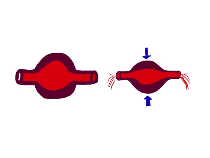
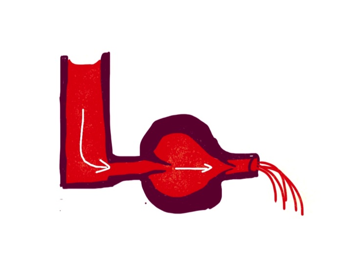
To make the blood go only in one direction, it is important to have valves at the inlet and outlet of the pump. There are three things helping to fill the pump when the muscle is relaxed. There needs to be a filling pressure, represented in the diagram below by the height of fluid in the left-hand reservoir. In the left side of the heart this is pulmonary venous pressure, in the right it is central venous pressure, most easily measured by looking at the height of blood in the neck veins. But lets first of all think about a simpler system with only one pump and one filling reservoir. Many years ago Frank and Starling discovered that if the filling pressure of a heart chamber affected how well it pumped blood out. The higher the filling pressure the stronger the contraction, making the pump work harder.
A lot of hollow things in our body work in a similar way. The uterus starts to contract when it gets stretched enough by the increasing size of the unborn baby. Our intestines work much harder if they are blocked, causing the wall to be stretched – this results in colic. The same is true for the gallbladder and ureters- both painfully contract when a stone causes a blockage and the muscular wall is stretched.
The second factor helping our heart chambers to fill up is suction. A turkey baster bulb will suck up fluid because the rubbery stuff it is made of wants to spring back to its normal shape. The same is true for the ventricles of the heart – they actively suck fluid in while relaxing during diastole. I am told that if you put an isolated animal heart into a bucket of oxygenated physiological saline solution it will move around like a squid – sucking liquid in and squirting it out to propel it along in the fluid. It could only do that if the ventricle actively sucks in fluid.
The third important factor in ventricular filling is the contraction of the atria. These give a bit of extra stretch to the ventricles by pumping in some more blood to help encourage them to pump it out a bit harder.
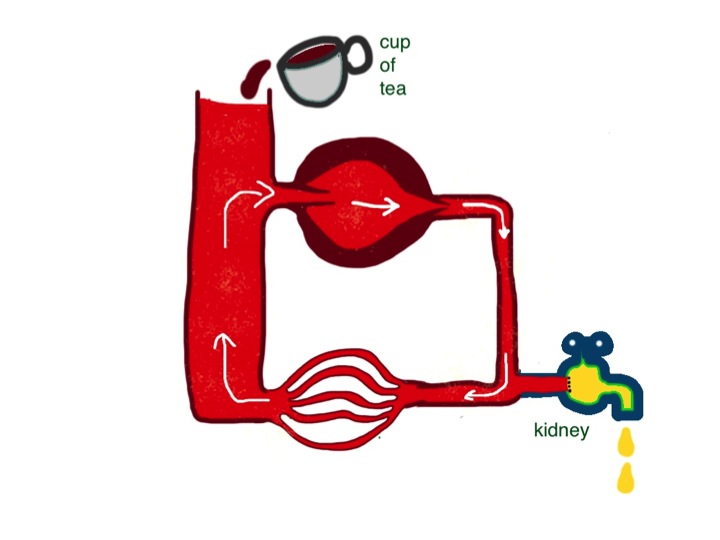
The heart pumps blood around a circuit – the circulation. It first goes into the aorta, but the vessels get progressively smaller in diameter, ending up with capillaries which have an internal diameter of less than that of a red blood cell – about 7 microns.

Pushing blood through the large vessels is very easy, but much more difficult through the smaller vessels. There is a strange relationship between the amount of resistance to flow and the internal diameter of a pipe which was discovered by Pouseuille- resistance is inversely proportional to the fourth power of the internal diameter. In practice this means that most of the resistance to flow is in small vessels with a hole down the middle measuring less than one tenth of a millimetre – these are also known as resistance vessels. Because of Pouseuille’s law, resistance vessels can constrict or relax a very small amount to cause a large change in flow to any particular organ – depending on its requirements for oxygen.
Back to the circuit. The arteries are thick-walled, with a relatively small hole down the middle. Veins are thin walled and can accommodate more volume. So most of the blood in our circulation is in the veins. Blood is pumped into the large arteries, is squeezed through the resistance vessels, and then ends up in the veins. You will not be surprised to know that the amount of pressure in the large arteries depends both on how hard the pump is pumping – cardiac output, – and how much resistance to flow there is in the small arteries – peripheral vascular resistance. Pressure = Flow x Resistance.
One thing our bodies really care about is the pressure in our large arteries – commonly known as blood pressure. It is continually monitored by pressure-sensing devices in the aorta and carotid arteries. These baroreceptors send messages to the brain stem (medulla). If the pressure is low, the medulla sends messages via the sympathetic nervous system to the heart to make it pump harder, and to the resistance vessels to make them constrict a bit to get the pressure back to normal. The medulla also sends messages to the veins to make them constrict – we will see why in a moment.
Looking at our simple circuit, blood pressure is determined by how hard the heart is pumping, and how much resistance to flow there is in small vessels. What determines the pressure on the venous side – the filling pressure of the reservoir (on the left side in the diagram)?

If the heart pumps less hard, or the resistance changes – I hope you can see that neither of these things will make a difference to filling pressure in the reservoir in this simple system. When students talk about “back pressure” they seem to forget that the blood goes round in a circuit. Failure of any part of the heart will not, on its own, affect filling pressure of the right side of the heart. Central venous pressure is determined by only two things: the amount of fluid in the system and how much veins are constricted. The amount of fluid in the circulation is affected by (a) how much fluid we drink every day and (b) how much we lose. Most of the fluid loss is in the form of urine (see previous post). It is the job of our kidneys to regulate the volume of blood in our circulation. Normally they do a fabulous job. If I drink a pint of beer, half an hour later I will be needing to have a pee – lots of dilute urine has been produced to get rid of all the extra water. If I eat two packets of crisps with the beer, over the next few hours my kidneys will get rid of the extra salt with no problem. Kidneys work in a timescale of hours.
Venous constriction works in a timescale of seconds. If I am in a chair and about to get up and walk around, my brain stem will send messages via the sympathetic nervous system to my veins to tell them to constrict. Squeezing this complex network of floppy tubes in my legs, belly and chest will increase cardiac filling pressure and, as Frank and Starling discovered, increase the force of contraction of my heart to supply my muscles with more oxygen. Understanding of how heart failure causes problems needs an understanding of how kidneys and veins work, not just the heart.
Now I am going to make the circulation model a bit more complicated. We have two pumps, not one. They are joined in series. The right heart pumps blood into the lungs. It returns to the left heart which pumps it round the rest of the body. The diagram shows the two pumps separated, but of course in real life they are part of the same organ.

The fact that they are arranged in series clearly could lead to problems. What if the right heart tries to pump more than the left? Why is this not normally a problem? The way the system is set up is that the right heart pumps blood through the lungs into the pulmonary veins. Here the pressure is higher than the normal 7cm water central venous pressure – in health about 16cm water. This higher pressure in the pulmonary vein is useful priming device so the left ventricle, which is bigger and stronger, can be rapidly filled and has plenty of pressure to stretch the rubbery myocardium to enable it to contract hard and generate adequate systemic arterial blood pressure. When right heart output increases, the pulmonary venous pressure will temporarily increase, and left ventricular output will in turn increase because of the Frank-Starling law, and subsequent reduction of pulmonary venous pressure to normal. The end result is that the output of the left side of the heart matches that of the right, and pulmonary venous pressure has not changed much – all sorted. This does mean, if you think about it, that the cardiac output is being controlled by the right heart – it pumps what it wants and the left heart has to follow suit. It is not always the case that the bigger, stronger partner is in control. John has found that out recently.
What controls right heart output? Well the brain stem has a big say, by constricting veins and increasing filling pressure and by sending messages to the heart via sympathetic nerves. But the kidneys are also really important – by altering fluid balance they can control filling pressure. Kidneys are involved in turning the dials and can really mess things up when they get it wrong, as we shall see.
Lets get back to John. His left ventricle was damaged by a sizeable myocardial infarction a few years ago (see chest pain and horsemeat lasagne below). Soon after had an echocardiogram which showed that the part of his left ventricle was not contracting as well as it should – the overall ventricular ejection fraction was found to be 44%. It was probably about 60% before. The rest of his left ventricle which was not damaged has to work harder to maintain his cardiac output and blood pressure. Heart muscle is different from ordinary skeletal muscle – the sort which makes our arms and legs work. Skeletal muscle responds very well to being worked hard. It grows bigger and stronger to more exercise we take. The heart muscle does this to an extent, but eventually it seems to give up and stop working so well. I don’t think anyone quite understands why this is. If experimental animals are infused with isoprenaline, a drug which makes the heart beat strongly by stimulating heart muscle cells in a similar way to noradrenaline released by sympathetic nerves, their hearts will fail in about 2 weeks. Beta blockers stop this sympathetic stimulation, and patients with heart failure live longer if they take them regularly.
Poor cardiac output stimulates the kidneys to produce renin. This is an enzyme which generates angiotensin I. Conversion of angiotensin I to angiotensin II on the surface of blood vessels helps restore blood pressure but also has a bad effect on heart muscle. It seems to make the muscle cells produce damaging free-radicals and oxidising agents which cause cardiac muscle death. ACE inhibitors prevent the formation of angiotensin II and protect the heart. But despite these drugs, once the ventricle is badly damaged, often the remaining heart muscle starts to fail after a period of time. Clearly in John’s case, another small myocardial infarction may have tipped him into symptomatic heart failure, despite the statins, omega-3 (fish oil), aspirin, and lack of steak and kidney pudding.
What happens when the left ventricle fails to pump properly? The right heart output is determined by central filling pressure and sympathetic activity. If it pumps blood through the lungs to the pulmonary veins, and if the left ventricle is not working, it will cause the pulmonary venous pressure to rise – lets say from the normal 12cm water to 20cm water. This rise in pressure will force the damaged left ventricle to pump harder until it can remove all the blood from the lungs that is being delivered. The right heart will not be too pleased with this- it is trying to pump what it thinks is the right amount of blood but is having a problem. As the pulmonary venous pressure rises, assuming the resistance to flow through the lungs does not change, the pressure in the pulmonary artery rises by the same amount as the rise in pulmonary venous pressure. The right ventricle will have to do more work to pump out blood into the lungs. Now, the left heart normally does not mind pumping at high pressure and doing lots of work – that’s what it is designed for. The right heart is just no good at it – it’s not lazy but it just does not have the muscle. (I put that bit in in case my wife decides to read this).
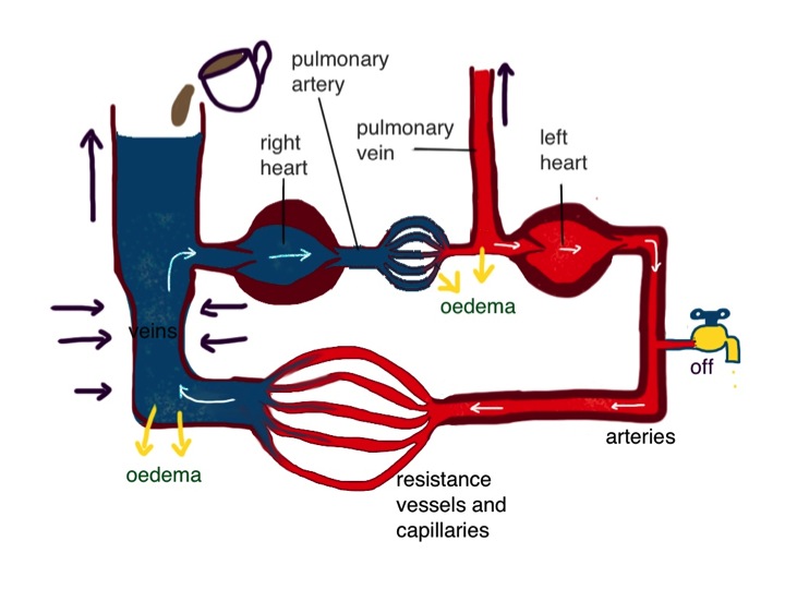
So, as a result the right heart can pump out less blood and we are left with a new balance – lower cardiac output and higher pulmonary venous pressure. The right heart is under strain because it is having to pump against higher pressure and the left heart is under strain because its filling pressure is higher than normal, making the undamaged parts also work harder than normal.
That’s fine until the kidneys and the brain get involved. They were doing a good job when John was healthy but in a crisis they make some bad management decisions. When cardiac output drops, blood pressure drops in proportion. Baroreceptors sense this and let the medulla know “Houston, we have a problem”. Increased sympathetic supply to small blood vessels is a good idea. Noradrenaline is released from nerves on the surface of blood vessels acting on alpha 1 receptors in the smooth muscle cell membrane. This causes the vessel to contract, making the holes down the middle smaller. Peripheral resistance increases and so does blood pressure. That was a good decision. There are similar sympathetic nerves which supply the heart. They are activated and the nerves again release noradrenaline, this time acting on beta receptors. The effect is to increase the rate and force of contraction of the heart, increasing cardiac output and blood pressure. Fine in theory, but this increase in work rate will increase cardiac oxygen consumption. John’s heart has a problem with getting enough oxygen because his coronary arteries have been ruined by too many steak and kidney puddings. Sympathetic stimulation can do more harm than good –that’s when reading the theory book goes wrong – a bad decision. And that is why we us beta blockers in patients with coronary artery disease and give them in patients who have had a myocardial infarction.
Another bad decision is to send messages to the veins to constrict. Increasing right heart filling pressure seems like a good idea. The right heart responds by pumping more blood into the lungs, not aware that the left heart is having a problem. If the pulmonary venous pressure goes above 25cm water there are serious problems. Fluid starts leaking out of the circulation in the lungs and causes pulmonary oedema. The fluid causes swelling of the gas-exchange membrane which allows oxygen to pass from the lungs to the blood, and for carbon dioxide to get out. Increased carbon dioxide concentration and reduced oxygen in the bloodstream are sensed by chemoreceptors in the carotid artery and the medulla (the respiratory centre which controls breathing is a close neighbour to the vasomotor centre which controls blood pressure). Houston panics. John (and his wife) also panics – he is fighting for breath. Houston is in panic mode – not only are its baroreceptors and chemoreceptors telling it that things are going wrong – messages are coming down from the White House – the cerebral cortex.
“More sympathetic stimulation” says Houston. Another bad decision. The increased sympathetic increases venous constriction and right heart filling. We are in a bad visious cycle, throwing petrol onto the flames. Just when Houston was beginning to give up Superwoman arrives. The nice ambulance woman has been trained not to panic – she’s seen it all before. It took her about 2 seconds to realise John had acute pulmonary oedema. She sat him up, gave him oxygen and got him to open his mouth and sprayed nitroglycerin under his tongue. Don’t worry dear- you’ll be fine in a moment. And he was. Reassurance works wonders in acute pulmonary oedema by reducing panic. Nitroglycerin or glyceryl trinitrate as we call it in the UK also works very well by selectively dilating veins, having less effect on the small resistance arteries. This reverses the bad vicious cycle, reducing right heart filling pressure, reducing right ventricular output and reducing pulmonary oedema, which reduces carbon dioxide levels in the blood which makes Houston much happier “was that a bird or a plane?”
I know of no evidence to support this, but I feel pretty sure the reason opiates like morphine work to reduce pulmonary oedema is to supress the respiratory centre and reduce central sympathetic output. Opiates certainly don’t work directly on veins to cause vasodilatation.
The problem with the medulla and normal cardiovascular reflexes is that they were designed to get us out of trouble when blood pressure falls from volume loss from trauma, diarrhoea or sepsis. Blood pressure drop from left ventricular failure is not in the manual. Primitive humans did not eat steak and kidney puddings.
The kidney is no better when it comes to heart failure. It knows there is a problem with cardiac output because it can sense it not getting enough blood. It makes the wrong assumption that this is due to circulating volume loss. The kidney’s response to low cardiac output is to stop producing urine. John is still drinking cups of tea and eating salty food, but he is not excreting it. The salt and water has to go somewhere – it increases the volume of blood in the circulation and increases right heart output, predisposing towards pulmonary oedema. The increase in blood volume can only go so far before it leaks out of the circulation and then equilibrates with extra-vascular interstitial fluid to produce ankle oedema. The only way to sort this out is to give diuretics like furosemide. This will force the reluctant kidneys to produce more urine and get the input/output balance right.
Many patients are fearful of taking daily furosemide, thinking that it will make them pass lots of urine all the time and make their lives difficult. Certainly, after the first few doses, daily urine volume increases, but it does not take much thinking about to realise that urine volume will soon settle down to be the same as the volume of fluid drunk each day. It can alter the pattern of urine output, so that for six hours after taking furosemide the volume is higher than normal, but for the next eighteen hours it is less. It might mean that patients need to get up at night to pass urine less often. Furosemide used to be called Lasix, because its effect lasts six hours.
I have explained why patients with heart failure benefit from nitroglycerin, morphine, beta blockers and furosemide. What about ACE inhibitors. As well as producing of free radicals in the heart, angiotensin II has an effect on veins. It does not directly constrict them, but enhances the effect of sympathetic activation originating in the medulla. That means that if the circulating levels of angiotensin II are very high, which is the case in heart failure, normal exercise will result in an exaggerated constriction of veins, an excessive rise in central venous filling pressure and too much blood being pumped into the lungs which the damaged left ventricle can’t handle. This will result in breathlessness. The main benefit patients with heart failure notice when they start ACE inhibitors is a reduction of breathlessness on exertion. The reduction of free radical production is a separate long-term benefit which will keep John alive longer.
So why do students (and doctors) talk about right heart failure as the cause of fluid retention and raised venous pressure when the damage is to the left side of the heart? I think the most likely reason is that in patients with true right heart failure, secondary to severe lung disease or pericardial tamponade, oedema can become really impressive. But again, the cause is not “back pressure”. If the right heart fails to pump properly, say the cardiac output drops from 5 litres/min to 2 litres/min, then the left heart can only pump out 2 litres/min. This means that the kidney again mistakenly goes into shutdown mode and fluid accumulates. The difference here is that pulmonary oedema is not going to be a problem, as the left heart is working normally, keeping pulmonary venous pressure normal. Under these circumstances patients sometimes present with gross oedema in their legs and fluid in their abdominal cavity, without presenting earlier with breathlessness. Thus historically, gross oedema is associated with right heart failure. Unless you can think of a better reason.
I can’t finish discussion of heart failure without mentioning preload and afterload. Anyone who mentions these terms when talking about humans, in general, is using the terms inappropriately. Preload and afterload were terms invented, I think, by Braunwald and Epstein. They were two brilliant cardiological physiologists who were interested in how filling pressure and arterial pressure affected cardiac ventricular function. They devised very clever experiments on isolated rat heart ventricular strips in organ baths. They wanted to look at the effect of increased filling pressure – so they put a spring in the organ bath to produce more tension and looked at how that affected contraction – they called it preload but never meant it to be anything but a proxy for filling pressure. Similarly, they thought “how can we model how the ventricle contracts against higher arterial pressure?” They cleverly added weights to be pulled up by the contracting ventricular strip – to make the ventricle do work. They called this afterload, but again did not mean it to be used to describe the in-vivo situation. If I ask one of the junior doctors “what is that patient’s preload?” there is no answer. They can tell me that the neck veins are distended. If I ask “what is that patient’s afterload?” they will rightly look at me strangely. But of course they can tell me what his blood pressure is.
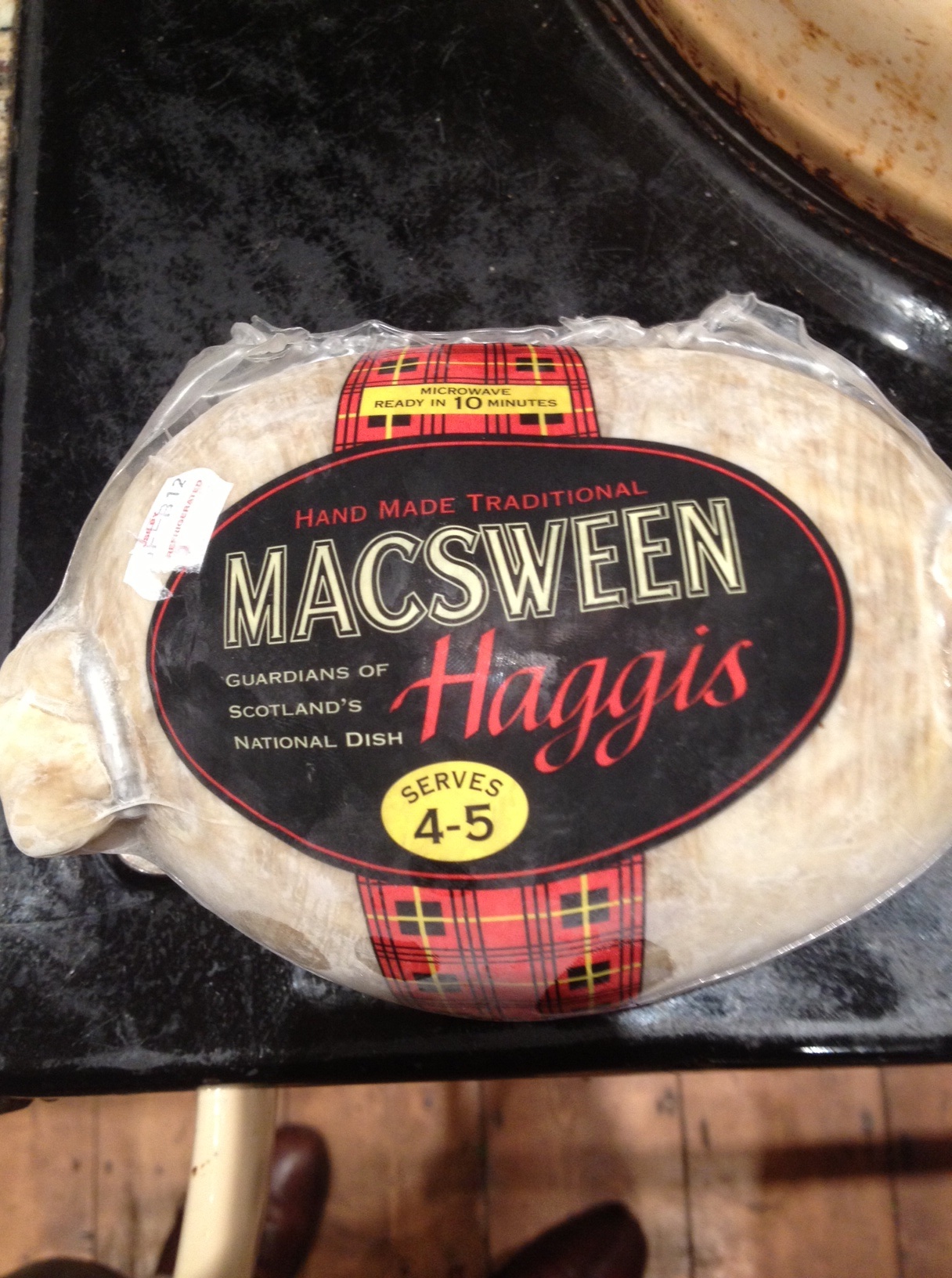
The food link this week is haggis. This is traditionally made from heart, lungs and kidney. Particularly appropriate for this discussion on heart failure. I like haggis roasted, not boiled. When roasted the skin, made from a sheep’s stomach, goes all brown and crispy. You must serve it with neeps (bashed swede, or rutabaga in the US) and tatties (mashed potato). Gravy is good but not necessary if you have a glass of whisky with it. Unfortunately I do suspect it is not much better than steak and kidney pudding for my coronary arteries as it also contains suet – the most saturated fat available. But it does have oatmeal.

Fascinating … thank goodness this is only of academic interest but what a morale boosting improvement it would be to call this condition almost anything other than ‘heart failure’.
Eye opening description!
Great “lecture” and interesting theory. Good reads…
Thanks Alexander
Not many manage to read it all. It is fairly hard going but hope you understood it
Dropsy